MRI Finds Breast Cancer Following Conservation Surgery
By MedImaging International staff writers
Posted on 04 Jul 2017
The results of a new study have compared the outcomes of two different combined screening methods for detecting new breast cancers in women after breast conservation surgery and radiotherapy.Posted on 04 Jul 2017
The researcher compared screening using both mammography and Magnetic Resonance Imaging (MRI), and screening using mammography and ultrasound, for patients whose breast cancer was first diagnosed at an age of 50 or less.

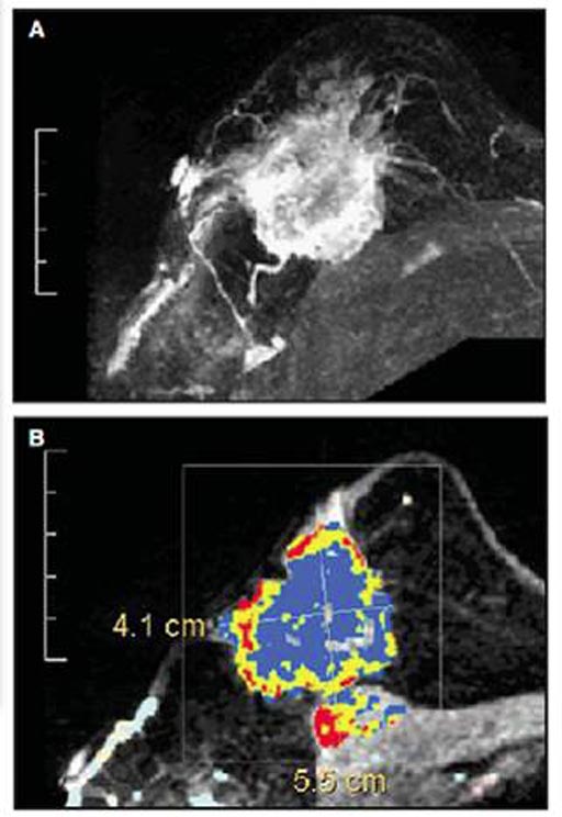
Image: Research shows MRI can detect breast cancer that is missed by mammography and ultrasound (Photo courtesy of the Seoul National University College of Medicine).
The results of the multi-center study in which 754 women were enrolled, were published online in the June 22, 2017, issue of the journal JAMA Oncology by researchers from the Seoul National University College of Medicine (Seoul, the Republic of Korea). The researchers performed annual mammography, breast MRI, and breast ultrasonography, for both contralateral (opposite) and conserved breasts during the three-year study.
During the study 17 cancers were diagnosed, of which 13 were stage 0 or stage 1 cancers. The researchers found that when MRI screening was used in addition to mammography, another 3.8 cancers per 1,000 women were discovered. When ultrasonography screening was added to mammography instead of MRI only 2.4 new cancers were detected.
The authors of the study, conclude, "After breast conservation therapy in women 50 years or younger, the addition of MRI to annual mammography screening improves detection of early-stage but biologically aggressive breast cancers at acceptable specificity [correctly identifying people who don’t have disease]. Results from this study can inform patient decision-making on screening methods after breast conservation therapy."
Related Links:
Seoul National University College of Medicine


