Acoustic Breast Imaging System Predicts Malignancy
By MedImaging International staff writers
Posted on 23 May 2017
A novel opto-acoustic (OA/US) system detects both tumor angiogenesis and the relative reduction in the oxygen content of blood that occur in cancer.Posted on 23 May 2017
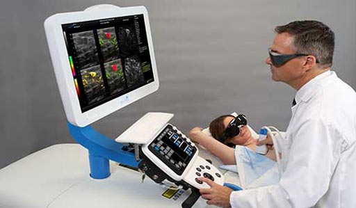
The Seno Medical Imagio OA/US breast imaging system combines both laser optics and conventional ultrasound technology to deliver fused functional and anatomical imaging of breast masses in real time. The images are generated based on hemoglobin concentration and oxygenation level, as the growth of malignant tumors is supported by angiogenesis to provide the necessary hemoglobin for that growth. The increased concentration of hemoglobin is a prime indicator of cancer.

Image: A new opto-acoustic system detects breast masses in real time (Photo courtesy of Seno Medical).
In contrast to existing optical and ultrasonic imaging techniques that create images by transmitting energy in one wave form and detecting that energy in the same form, OA/US imaging transmits light energy but detects sound energy; by using multiple colors of laser light a much broader range of data can be captured, which makes the functional images possible. The images are also generated without using ionizing radiation or injection of contrast agents. The Seno Medical Imagio system has received the European Community CE mark of approval.
In a recent Phase III pilot study to determine the histopathologic basis of OA/US breast imaging, 92 patients with 94 solid or complex cystic and solid breast masses assessed with conventional diagnostic ultrasound (CDU) were also imaged with OA/US. The mean OA scores were higher for malignant masses compared to benign masses for all individual internal and external features, as well as for combined internal and external OA features. The study was presented at the American Roentgen Ray Society (ARRA) annual meeting, held during May 2017 in New Orleans (LA, USA).
“Opto-acoustic imaging provides real-time anatomic and functional information without ionizing radiation or the need for IV contrast injection, making it a potentially safer and more convenient option for patients,” said lead author and study presenter Reni Butler, MD, of Yale University School of Medicine. “This could help us decrease the number of unnecessary breast biopsies performed for benign findings, reducing patient anxiety, discomfort, and healthcare cost.”


