Carotid Ultrasound Examinations Help Identify Stroke Risk
By MedImaging International staff writers
Posted on 06 Dec 2015
A new study concludes that carotid ultrasound is highly accurate in detecting the presence of calcified atherosclerotic lesions larger than 8 mm3, but less accurate in detecting smaller volume calcified plaques. Posted on 06 Dec 2015
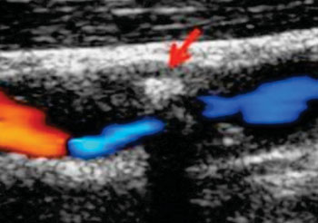
Researchers at Umea University (Sweden) conducted a study that analyzed 94 carotid arteries from 88 patients (mean age 70 years, 33% females) to determine the accuracy of ultrasound B mode in detecting atherosclerotic calcifications during pre-endarterectomy examinations. After the procedure, the excised plaques were quantified using cone beam computed tomography (CBCT), from which the calcification volume (in mm3) was calculated. Carotid artery calcification by the two imaging techniques was then compared using conventional correlations.

Image: Preoperative carotid ultrasound evaluation (Photo courtesy of Umea University).
The results showed that carotid ultrasound was highly accurate in detecting calcifications, with a sensitivity of 88.2%. Based on calcification volumes as measured by CBCT, the researchers divided the plaque into four groups: less than 8; 8–35; 36–70; and over70 mm3. They found that calcification volumes larger than 8 mm3 were accurately detectable by ultrasound, with a sensitivity of 96%. Of the 21 plaques with a smaller than 8 mm3 calcification volume; only 13 were detected by ultrasound, resulting in a sensitivity of 62%. The study was published in the August 2015 issue of International Journal of Molecular Sciences.
“We know that preventive surgical treatment of carotid stenosis is only beneficial for a small subgroup, and that most asymptomatic patients will do better with only medical therapy,” said lead author Fisnik Jashari, MSc, a doctoral student in the department of public health and clinical medicine. “By using ultrasound, we can identify the patients who are at a higher risk of stroke and thus would benefit from surgery. But preventing unnecessary surgical intervention in most cases is equally important.”
Historically, carotid calcification is detected by X-ray imaging. With the development of ultrasound examination of vascular pathology, carotid two-dimensional (2D) grey scale has become a routine investigation in vascular laboratories; but while ultrasound may detect the presence of calcification, it is an accurate means for quantifying its extent, unlike CBCT, which can detect calcification volumes as small as one mm3. However, any CT investigation carries the risk of significant radiation, as well as limited availability.
Related Links:
Umea University


