Ultrasound Provides Insights into Abdominal Aortic Aneurysms
By MedImaging International staff writers
Posted on 28 Oct 2014
Researchers are assessing the effectiveness of the use of ultrasound to study lethal abdominal aortic aneurysms (AAAs), a bulging of the aorta that is typically fatal when it ruptures, and for which there is no effective medical treatment.Posted on 28 Oct 2014
Abdominal aortic aneurysms are the 13th leading cause of death in the United States, killing about 15,000 yearly. “Most of the time people don’t even know it’s there, but if it does rupture they have internal bleeding from the largest artery in the body,” said Craig Goergen, an assistant professor in Purdue University’s (West Lafayette, IN, USA) Weldon School of Biomedical Engineering. “Eighty percent of the time the patient dies before getting to the hospital.”

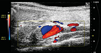
Image: Purdue University researchers are using ultrasound images like this one to study abdominal aortic aneurysms, a potentially fatal condition that is the 13th leading cause of death in the United States (Photo courtesy of Purdue University/Weldon School of Biomedical Engineering).
Researchers have used a technique called high-frequency small animal ultrasound to measure alterations in the progression of abdominal aortic aneurysms in lab animals. By learning more about aneurysms at the molecular level, researchers might be able to develop drug or stem cell therapies to keep them from growing larger, according to Dr. Goergen.
New findings were presented on October 25, 2014, by doctoral student Evan Phillips during the Biomedical Engineering Society’s annual meeting in San Antonio (TX, USA). The researchers are able to monitor small changes in the vessel as the aneurysm forms. Ultrasound imaging allows for visualization of the arterial layers that frequently separate from each other while the vessel grows. “The development of these aneurysms is a multifaceted process that has not been fully characterized, and no therapeutics have proven to effectively prevent aneurysm progression,” Dr. Goergen said. “What we want to do is understand what causes aneurysms to form and grow so that we can then develop better strategies to prevent that from happening.”
The ultrasound technique also uses color Doppler imaging, making it possible for researchers to collect color-coded blood flow data showing the direction and speed of circulation, both of which are factors that might influence how aneurysms develop and expand. The frequency of a wave changes as it moves toward or away from the observer, a phenomenon known as the Doppler effect. Color Doppler images reveal red flow moving toward the ultrasound transducer and blue flow moving away. The efforts are laying a basis for additional studies that might use these strategies in a clinical setting or to investigate potential therapeutics, according to Dr. Goergen.
Human aortic aneurysms typically begin at 3–4 cm in diameter and are located in the abdomen below the kidneys. The aorta extends from the heart through the body and ultimately branches into two smaller arteries leading to the legs. Most at risk are older men who have high blood pressure and a history of smoking. Men over 65 who fit this profile are advised to contact their doctor about ultrasound screening to determine if they have an aneurysm.
“Unless you feel a pulsating mass, develop unusual back pain, or are screened with ultrasound, most people don’t even discover that they have it. If you are diagnosed, most surgeons won’t treat aneurysms less than 4 cm. Unfortunately, there is no medicine that can prevent aneurysms from growing,” Dr. Goergen said. “Doctors can give you statins to lower your cholesterol levels or anti-hypertension medication to lower your blood pressure, but there are no medications that can directly prevent aneurysm expansion.”
However, researchers can analyze laboratory animals that mimic conditions in humans to provide insights into the mechanisms behind aneurysms. The group led by Dr. Goergen used ultrasound imaging to quantify changes over a four-week period as aneurysms developed and expanded in mice and rats. “We’re doing three-dimensional ultrasound that can reconstruct that whole aorta,” Dr. Goergen said.
Because this technique is noninvasive, it may ultimately be used on humans to diagnose and monitor aneurysm progression.
Related Links:
Purdue University


