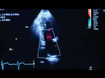
Mobile, Cardiovascular Ultrasound Features Stress Echo and Transesophageal Echocardiography Capabilities
By MedImaging International staff writers
Posted on 14 Jul 2014
A 58.5-kg mobile cardiovascular ultrasound system features innovative quantitative features such as stress echo, and transesophageal echocardiography (TEE) capabilities, designed for healthcare providers looking for a cost-effective echo technology. Posted on 14 Jul 2014
GE Healthcare (Chalfont St. Giles, UK) recently reported on the US Food and Drug Administration (FDA) clearance for its new Vivid T8 cardiovascular ultrasound device. The Vivid T8 system has been optimized by combining the established cardiac imaging capabilities of GE Vivid systems with exceptional shared services performance of the company’s LOGIQ systems. The Vivid T8, cardiovascular ultrasound system is rugged, effective, and full of features, yet still cost-effective and convenient to use.

Image: The Vivid T8 cardiovascular ultrasound system offers quantitative features such as stress echo and transesophageal echocardiography (TEE) capabilities (Photo courtesy of GE Healthcare).
“Performance, complete echocardiography features, shared services and reliability were the key priorities in developing the Vivid T8,” said Al Lojewski, GM of cardiovascular ultrasound for GE Healthcare. “In developing the system, we put it through more than 20 hours of intense vibration, shock, and thermal testing to ensure its reliability in the settings outlined by our customers. The result is a value system unlike any other we’ve commercialized at GE. With the growing global crisis of cardiovascular disease, we wanted to develop an affordable, reliable solution that could potentially allow clinicians around the world increased access to high performance ultrasound.”
The Vivid T8 is a mobile system intended for use in a range of conventional as well as harsh, demanding environments. It is sturdy enough to the challenges of even the busiest ultrasound imaging practices and clinical settings and is backed with three years of service coverage.
Designed to be easy to use yet clinically versatile, the T8 features an intuitive user interface with touch-screen capabilities, rotary dials, and patient management buttons with the look and feel of a true Vivid console. Furthermore, it provides access to up to four transducer ports allowing increased, easier access to more clinical procedures.
The system allows healthcare providers access to sophisticated quantitative tools and delivers excellent shared service image quality, with options to tailor the system to fit each healthcare provider’s needs.
Key clinical applications include: (1) Cardiac tissue velocity imaging, which captures dynamic information from moving heart tissue to quantify left ventricular function. (2) The AutoEF function automatically evaluates left ventricular ejection fraction (EF) using an automated, speckle-tracking region of interest (ROI) tool. (3) The SmartStress feature automatically adjusts settings to help enhance workflow, reproducibility, and diagnostic confidence. (4) Automated function imaging assesses and quantifies left ventricular wall motion at rest, and calculates parameters to describe left ventricular wall function. (4) The Auto IMT provides automatic edge detection for intima-media thickness (IMT) and auto-completes required measurements. (5) Adult multiplane transesophageal echo transducers are available for specific echo examinations. (6) Monitoring and guidance to support interventional procedures; (7) Virtual convex extends the field-of-view (FOV) when using linear transducers; (8) the LOGIQ view increases the field of view to image large organs that typically cannot be seen in a single image; (9) The B-Flow function provides advanced spatial and temporal resolution to help assess blood flow and vessel wall structure without the limitations of Doppler. (10) Lastly, blood flow imaging helps enhance visualization of blood flow dynamics using a unique signal-processing algorithm.
Related Links:
GE Healthcare


.jpg)
