New Ultrasound Device May Help to Detect Risk for Stroke and Heart Attacks
By MedImaging International staff writers
Posted on 06 May 2014
New prototype ultrasound technology could help detect arterial plaque that is at high risk of breaking off and causing a heart attack or stroke.Posted on 06 May 2014
Plaque around the heart accumulates in arteries as people get older. Some types of plaque are considered to be “vulnerable,” meaning that they are more likely to detach from the artery wall and cause heart attack or stroke.
.")
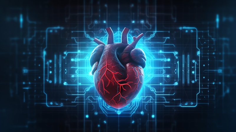
Image: The new ultrasound device will help identify vulnerable plaque that increases risk of heart attack or stroke (Photo courtesy of Xiaoning Jiang).
Researchers from North Carolina (NC) State University (Raleigh, USA) and the University of North Carolina at Chapel Hill (USA) have developed the ultrasound device. “Existing state-of-the-art technologies are capable of determining if plaque is present in the arteries, but can’t tell whether it’s vulnerable. And that makes it difficult to assess a patient’s risk,” says Dr. Paul Dayton, coauthor of a paper on the new device and professor in the joint biomedical engineering department at NC State and Chapel Hill. “Our goal was to develop something that could effectively identify which plaques are vulnerable.”
There are two ultrasound techniques that can help detect vulnerable plaques, but both make use of contrast agents called “microbubbles.” The first technique is to identify vasa vasorum in arteries, which are clusters of small blood vessels that frequently infiltrate arterial plaque, and which are considered indicators that a plaque is vulnerable. When microbubbles are injected into an artery, they move with the blood flow. If vasa vasorum are present, the microbubbles will flow through these blood vessels as well, effectively highlighting them on ultrasound images.
The second technique is called molecular imaging, and relies on the use of “targeted” microbubbles. These microbubbles fasten themselves to specific molecules that are more likely to be found in vulnerable plaques, making the plaques emphasized on ultrasound images.
“The problem is that existing intravascular ultrasound technology does not do a very good job in detecting contrast agents,” stated Dr. Xiaoning Jiang, an NC State associate professor of mechanical and aerospace engineering, an adjunct professor of biomedical engineering and coauthor of the article. “So we’ve developed a dual-frequency intravascular ultrasound transducer which transmits and receives acoustic signals. Operating on two frequencies allows us to do everything the existing intravascular ultrasound devices can do, but also makes it much easier for us to detect the contrast agents—or microbubbles—used for molecular imaging and vasa vasorum detection.”
The prototype device has performed well in laboratory testing; however, the researchers reported that they are still enhancing the technology. They plan to establish preclinical studies in the near future.
The study was published in the May 2014 issue of the journal IEEE Transactions on Ultrasonics, Ferroelectrics, and Frequency Control.
Related Links:
North Carolina State University
University of North Carolina at Chapel Hill

Gold Member
Solid State Kv/Dose Multi-Sensor
AGMS-DM+

New
Ceiling-Mounted Digital Radiography System
Radiography 5000 C

Color Doppler Ultrasound System
DRE Crystal 4PX

Ultrasound Software
UltraExtend NX
.jpg)