Prostate Cancer Patient Receives First Treatment of Real-Time Brachytherapy Planning System
By MedImaging International staff writers
Posted on 29 Apr 2013
A prostate cancer patient is the first individual in the world to be treated using the latest generation of a real-time planning system for planning and performing cutting-edge high-dose-rate (HDR), ultrasound-guided brachytherapy treatments. Posted on 29 Apr 2013
The treatment was performed at the Levine Cancer Institute (Charlotte, NC, USA). “Our team was very impressed overall with this treatment planning system,” stated Dr. Michael Haake, chief, division of radiation oncology at Carolinas Medical Center, Levine Cancer Institute. “The total anesthesia time for the patient was just three hours, which is short for this type of treatment. He was discharged about two and half hours after he woke up.”

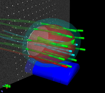
Image: The Vitesse software reconstructs needles in real time using 3D ultrasound data (Photo courtesy of Varian Medical Systems).
HDR brachytherapy involves delivering radiotherapy from inside the body by briefly placing a tiny radioactive source directly into the tumor or other targeted area. Using a robotic device called an afterloader, clinicians place the radioactive source into positions through needles that have been inserted into the region being treated. Under computer control, the source is then moved within the needles to create the specified dose distribution within the patient’s anatomy.
Using the latest version of Vitesse, developed by Varian (Palo Alto, CA, USA), HDR brachytherapy treatment plans can be created in a real-time environment, using ultrasound images generated in the operating room rather than CT scans generated postoperatively elsewhere. This avoids the need to move the patient to a CT scanner for imaging after the needles have been put in place. The process can now be completed completely within the Vitesse program, from capturing the ultrasound image to finalizing an approved treatment plan.
“From a practical point of view, the Vitesse software enabled us to monitor the location of the implant needle tips better in real time, by moving the needle as my dosimetry personnel mapped their position, which allowed us to actually leave the procedure room with a great treatment plan,” added Dr. Haake. “The real time planning allowed us to edit the plan during treatment, which would have been much more difficult and time consuming using our previous planning system.”
“This version of Vitesse reduces the number and complexity of steps involved in planning and completing a treatment,” said Tim Clark, marketing manager for Varian brachytherapy. “It eliminates the need for a data transfer to another software program, and avoids moving the patient to a CT scanner for images in the middle of the procedure. For these reasons, most clinicians will see a reduction in the amount of time needed to complete these treatments, often by as much as an hour and a half.”
Levine Cancer Institute is part of the Carolinas HealthCare System (CHS), which comprises 32 affiliated hospitals in North and South Carolina.
Related Links:
Varian
Levine Cancer Institute


 Guided Devices.jpg)


