Contrast-Enhanced Ultrasound Detects High-Grade Prostate Tumors Better with Fewer Biopsies
By MedImaging International staff writers
Posted on 09 Oct 2012
Contrast-enhanced ultrasound can detect high-grade prostate cancer better than established methods, making it a more suitable approach for screening clinically significant tumors and monitoring low-risk ones with fewer biopsies.Posted on 09 Oct 2012
Researchers from Thomas Jefferson University and Hospitals (Philadelphia, PA, USA) presented their findings from a phase III study online September 2012 in the Journal of Urology. The randomized, double-blind trial results revealed the technique, which uses microbubbles to track change in blood flow, revealed nearly three times as many higher grade tumors using half as many needle biopsies compared to systematic biopsy techniques.
“Today, a physician may sample 12 to 18 tissue cores from the prostate in order to help diagnose a patient. But with contrast-enhanced, that number drops to six or even less,” says lead author Ethan Halpern, MD, codirector of the Prostate Diagnostic Center Thomas Jefferson University Hospital and professor of radiology and urology at Thomas Jefferson University. “So it’s less invasive, and a more effective guidance tool. We’ve found that with contrast-enhanced ultrasound, we are much more likely to detect cancers on the image, and in this case, the higher grades.”
The clinical trial’s findings of 311 men, 118 of which had positive prostate cancer biopsies, showed that targeted biopsies using contrast-enhanced ultrasound with microbubbles detected significantly more higher volume/grade prostate tumors in men (55%) compared to a traditional prostate biopsy technique (17%).
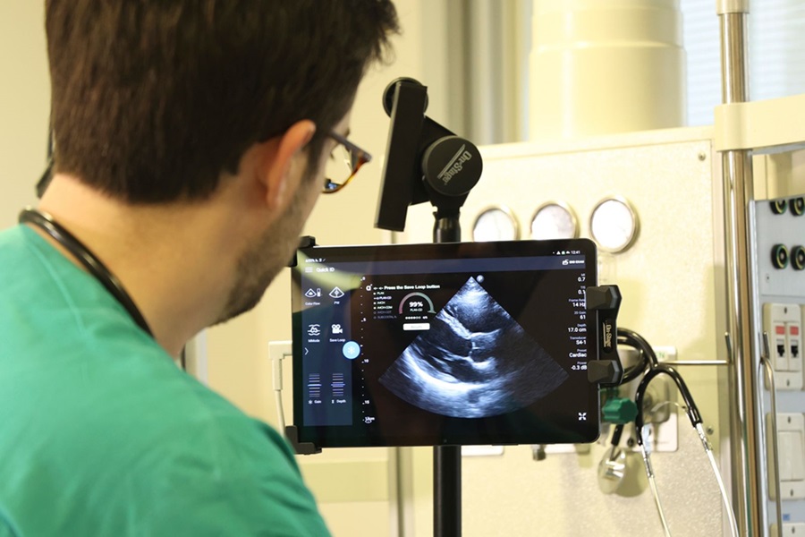
Ultrasound imaging of the prostate is typically used to evaluate the size of the gland and for needle placement during systematic biopsy, but is hindered by difficulty in distinguishing benign from malignant tissue. What makes contrast-enhanced ultrasound unique is the microbubble contrast agents, tiny bubbles of gas contained within a supporting shell that are injected into the patient to help better measure alterations in blood flow.
Prostate cancer, similar to many cancers, harbors abnormal blood vessel flow. This change in flow in the prostate can be measured by ultrasound; the microbubbles enhance the reflection of those ultrasound waves.
The technique has been effectively used in Europe for some time, but researchers at Jefferson reported it is ready for use in the United States. The US Food and Drug Administration (FDA) has not approved it for use for prostate screening, although it is used in other imaging applications.
In the clinical trial, researchers performed both targeted biopsies using contrast-enhanced ultrasound with flash replenishment maximum intensity projection microflow imaging on all patients, and a systematic 12-core biopsy protocol for comparison. The mean age of the patients was 62 years and a prostate-specific antigen (PSA) level of 6.5 ng/ml.
“Our ultimate goal is to perform a limited number of targeted biopsies and leave the rest of the prostate alone,” says Dr. Halpern. “This will provide a safer, more cost-effective approach to diagnosing prostate cancer.”
Study participants were also randomized to pretreatment with dutasteride, a drug used to treat an enlarged prostate, and placebo; however, no was significant difference in the proportion of positive biopsies for prostate cancer.
Dr. Halpern, who is lead investigator on the four-year, US National Cancer Institute (Bethesda, MD, USA)-supported trial, has been developing and modifying techniques to improve targeted biopsy of the prostate for more than 10 years, along with his colleagues at Jefferson, including Edouard J. Trabulsi, MD, codirector of the Prostate Diagnostic Center and associate professor of urology.
“It stands to reason that the cost-benefit ratio for prostate cancer screening will improve if PSA screening is followed by a limited targeted biopsy based on contrast-enhanced ultrasound,” concluded Dr. Trabulsi. “This also means contrast-enhanced ultrasound can act as another monitoring tool for active surveillance in low-grade cancer patients, potentially preventing unwarranted treatments.”
Related Links:
Thomas Jefferson University and Hospitals

Gold Member
Solid State Kv/Dose Multi-Sensor
AGMS-DM+

Thyroid Shield
Standard Thyroid Shield

New
Ultrasound System
Voluson Signature 18

New
CT Phantom
CIRS Model 610 AAPM CT Performance Phantom
.jpg)