Physician Interpretation Time Drastically Reduced by Automated Breast Ultrasound
By MedImaging International staff writers
Posted on 23 May 2012
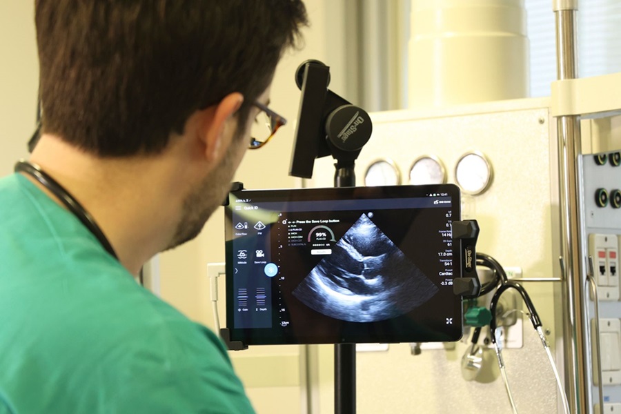
Automated breast ultrasound takes an average three minutes of a physician’s time, allowing for faster and more complete breast cancer screening of asymptomatic women with dense breast tissue, a new study revealed. Posted on 23 May 2012
Mammography misses more than one-third of tumors in women with dense breasts, according to Rachel Brem, MD, lead author of the study. “Ultrasound can and does detect additional, clinically significant, invasive, node negative breast cancers, that are not seen on mammography, but a hand-held ultrasound screening exam requires 20-30 minutes of physician time. Having a technique that takes just three minutes is a “game-changer” in appropriately screening these women,” said Dr. Brem.
The study, conducted at George Washington University Medical School (Washington DC, USA), quantitatively evaluated the time it took for radiologists to interpret automated breast ultrasound examinations. The average reading time for the three radiologists in the study was 173.4 seconds, said Dr. Brem.
Currently, automated breast ultrasound is limited in use, although a US Food and Drug Administration (FDA) panel just recently voted in favor of its efficacy and safety. “When automated breast ultrasound is integrated in the screening environment, we will see the detection of smaller, more curable breast cancers. The days of one size fits all approach to breast screening are passing.
Automated breast ultrasound provides us with a tailored approach based on the individual woman’s breast density,” Dr. Brem said. “When the Food and Drug Administration clears automated breast ultrasound for screening, I’m confident we will see a rapid integration of this approach into practice to improve cancer detection in women with dense breasts,” she said.
The study was presented May 5, 2012, at the American Roentgen Ray Society annual meeting in Vancouver (BC, Canada).
Related Links:
George Washington University Medical School

Gold Member
Solid State Kv/Dose Multi-Sensor
AGMS-DM+

Thyroid Shield
Standard Thyroid Shield

Ultrasound Needle Guide
Ultra-Pro II

New
Digital Radiography Generator
meX+20BT lite
.jpg)