Versatility, Portability, and Improved Image Quality All Key to Ultrasound Growth
By MedImaging International staff writers
Posted on 02 Aug 2011
An interview with GE Healthcare's Michael StockhammerPosted on 02 Aug 2011
Medical imaging technology has come a long way, according to market insiders, in particular, ultrasound. Since its development in the early 1950s, medical ultrasound has gained a major share of the imaging market next to X-ray. Today, ultrasound systems are ubiquitous, found typically in the medical environments of radiologists, obstetricians, cardiologists, and others. The technology is suitable for use in a wide range of patients, and is recognized as safe and patient-friendly.
Applications in new segments such as emergency medicine and the development of portable or hand-held units are driving clinical demand for these devices. Hospitals and various other healthcare institutions worldwide have adopted this modality, generating significant market prospects for manufacturers of ultrasound equipment. Ultrasound remains an effective and low-cost imaging technology.
Whereas the adoption of ultrasound technology was considerably faster in North America and Western Europe, an increasing confidence in portable systems has made its way to other regions of the world, such as Eastern Europe, Latin America, and parts of Asia-Pacific. Demand is coming from conventional applications, such as ob/gyn, and increasingly from point-of-care applications. The market research company InMedica (Wellingborough, UK) recently pointed out that the economic downturn is favoring the lower end of the cart-based ultrasound equipment segment. Furthermore, the US hand-held ultrasound segment is expected to exceed US$1.2 billion by 2016 due to technologic advances and improvements in three and four-dimensional (4D) capabilities, according to a report by market research firm iData Research (Vancouver, BC, Canada).
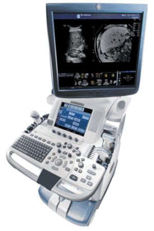
Success or failure in medical imaging primarily depends on image quality, emphasized Michael Stockhammer, EMEA (Europe, Middle East, and Africa) general manager of the ultrasound business unit of GE Healthcare (Chalfont St. Giles, UK), in an interview with Dr. Jutta Ciolek, regional director of Medical Imaging international. Stockhammer described GE’s progress in the field: “Interestingly, it took us a while to find our way into the radiology market. The actual breakthrough was the release of LOGIQ E9 ultrasound system two years ago. It grew to be the most successful ultrasound product in the entire ultrasound portfolio. Until the end of last year, we delivered about 3,000 systems worldwide – about 800 in Europe.”
The GE Healthcare executive went on to expand on the high-end aspects of the LOGIQ E9: “There’s a complete set of advanced tools, which we list under Expert Tools. This starts with features such as contrast-ultrasound, where you inject a contrast agent –primarily in the field of abdominal examination, i.e., liver, kidneys, pancreas – to get a better detection of malignant or benign lesions, but primarily to be able to classify these lesions. With the LOGIQ E9, we surely have an outstanding device for this, which made us acknowledged as one of the leading suppliers. We also added elastography capabilities to the LOGIQ E9 ultrasound platform.
Elastography uses mechanical compression to analyze the stiffness of tissues; the software calculates the strain in the region of interest after compression. This calculation creates an elastogram, which is a color overlay on top of the B-mode image that represents tissue elasticity. Pathologic lesions are typically considerably more resistant to compression than healthy tissue. A quality indicator monitors the operator’s compression technique. The LOGIQ E9 incorporates several advanced technologies, including the E series transducers, which feature significant improvements in sensitivity, bandwidth, and axial resolution and penetration.
Moreover, since the release of LOGIQ E9, we have a feature called Image Fusion, which enables us to use images from other modalities besides ultrasound for imaging, modalities like CT [computed tomography], MR [magnetic resonance], and soon PET [positron emission tomography]/CT, which can be imported into the device. We not only import pure two-dimensional data sets but entire volumes. These correlate points in the MR or CT dataset with ultrasound. And, as soon as you move the probe, the position of the probe over the magnetic field gets registered and shows you the corresponding layer in the CT and MR data sets.”
Another and arguably even more interesting aspect of the latest imaging advances, lies in the field of interventional radiology. According to Stockhammer, “if you think about conducting complex punctures as is often done today with CT, this is very cost-, and radiation-intensive, and blocks the device for some hours. If it were possible to redirect some of these examinations to ultrasound – and primarily decrease the time of the examination – it would have a massive positive cost-effect and quality for the patient.” He noted that with the biopsy guidance tool, the needle tip is referenced to a sensor attached to the ultrasound probe so that the path to a target can be determined and visualized before the skin is penetrated. “With tools such as biopsy guidance where you have the biopsy channel displayed on the screen – because a thin biopsy needle tends to bend while entering the tissue – you really track the actual position and hit the lesion exactly. And, this certainly is of great interest.”
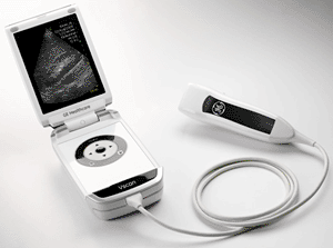
The GE Healthcare executive also commented on the Vscan, the flip-phone-like, ultra-portable ultrasound scanner, “This is our hope, our dream. It will still take some time. We are not the first company that has tried launching a portable ultrasound device; there were others before us, but with somewhat limited image quality. However, the technology never has been this advanced. The Vscan is the first product that in fact offers very good image quality coupled with Color Doppler -sensitivity in the fields of cardiology, abdominal sonography, anesthesia, as well as obstetrics. Actually, we label it as a “pocket-sized visualization tool.” This is because we deliberately want to make the customer aware that the device has its limitations and hence serves a different purpose when compared to a full-blown ultrasound-imaging device. It is not going to replace the ultrasound console or laptop ultrasound device anytime soon. But the device has been on the market for the past 10 months.” As to the clinical benefits of Vscan and its target users, Stockhammer added, “This is also something where we want to show that the quality of the examination in the field of 2D and in the field of color-echocardiography is very near the big console devices.”
Regarding where ultrasound technology is growing the strongest, the GE Healthcare executive commented, “first of all in Europe, which however primarily is a replacement market characterized by cut-throat competition, with very few new investments. Where we see the highest potential for growth is the field of point-of-care. These are new groups of customers that use ultrasound: from the fields of regional anesthesia, rheumatology, venous therapy, and physiotherapy. This is where two-digit growth figures can be seen, and we are massively active in this field. Not only with the development of dedicated devices –with a simplified interface in order to facilitate faster adoption time – and of course, the same as with Vscan, this offer has to be combined with training programs. This is what we promote.”
Related Links:
GE Healthcare


