Risk of Osteoporosis May Be Predicted by Ultrasound Exam
By MedImaging staff writers
Posted on 28 Jul 2008
Posted on 28 Jul 2008

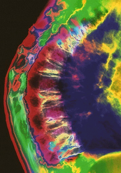
Image: Computer-enhanced X-ray of the curved and deformed thoracic spine of a woman suffering from osteoporosis (Photo courtesy of Mehau Kulyk).
The study, published in the July 2008, issue of the journal Radiology, reported that combined with certain risk factors, including a recent fall, or age, noninvasive ultrasound of the heel may be used to better select women who need further bone density testing, such as a dual-energy X-ray absorptiometry (DXA) exam.
"Osteoporosis is a major public health issue expected to increase in association with worldwide aging of the population,” said the study's lead author Idris Guessous, M.D., senior research fellow in the department of internal medicine at Lausanne University Hospital (Switzerland; www.chuv.ch). "The incidence of osteoporosis will outpace economic resources, and the development of strategies to better identify women who need to be tested is crucial.”
Osteoporosis is a disease that is characterized by low bone mass and the deterioration of bone tissue and is a major public health problem. According to the U.S. National Osteoporosis Foundation, 10 million Americans currently have osteoporosis and approximately 34 million more are estimated to have low bone mass, increasing their risk of developing the disease. Eighty percent of those affected are women.
"Patients with osteoporosis are not optimally treated because of a lack of general awareness,” Dr. Guessous said. "A simple prediction rule might be a useful clinical tool for healthcare providers to optimize osteoporosis screening.”
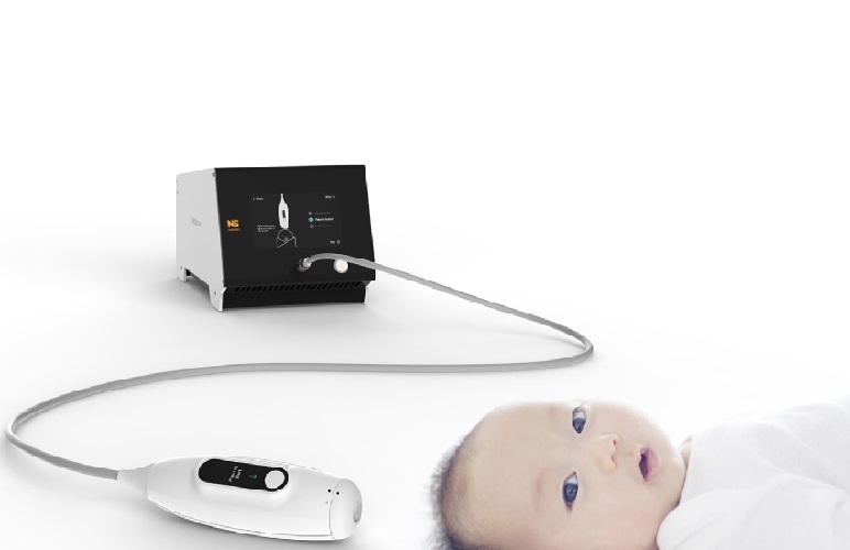
In the three-year multicenter study, 6,174 women age 70 to 85 with no previous formal diagnosis of osteoporosis were screened with heel-bone quantitative ultrasound (QUS), a diagnostic test used to evaluate bone density. QUS was used to calculate the stiffness index, which is an indicator of bone strength, at the heel. Researchers added in risk factors such as age, history of fractures, or a recent fall to the results of the heel-bone ultrasound to develop a predictive rule to estimate the risk of fractures. The findings demonstrated that 1,464 women (23.7%) were considered lower risk and 4,710 (76.3%) were considered higher risk.
Study participants where mailed questionnaires every six months for up to 32 months to record any changes in medical conditions, including illness, changes in medications, or any fracture. If a fracture had occurred, the patients were asked to specify the fracture's precise location and trauma level and to include a medical report from the physician in charge.
In the group of higher risk women, 290 (6.1%) developed fractures whereas only 27 (1.8%) of the women in the lower risk group developed fractures. Among the 66 women who developed a hip fracture, 60 (90%) were in the higher risk group.
The results revealed that heel QUS is not only effective at identifying high-risk patients who should receive further testing, but also may be helpful in identifying patients for whom further testing can be avoided. "Heel QUS in conjunction with clinical risk factors can be used to identify a population at a very low fracture probability in which no further diagnostic evaluation may be necessary,” Dr. Guessous said.
Related Links:
Lausanne University Hospital


