Simple Brain Scan Diagnoses Parkinson's Disease Years Before It Becomes Untreatable
Posted on 19 May 2025
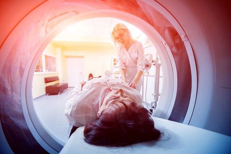
Parkinson's disease (PD) remains a challenging condition to treat, with no known cure. Though therapies have improved over time, and ongoing research focuses on methods to slow or alter the disease’s progression, early diagnosis is critical. Detecting the disease early would allow for treatments to be administered when they have the highest potential to preserve brain function. In a groundbreaking study, an international team of researchers has demonstrated that it may soon be possible to diagnose PD years in advance using functional magnetic resonance imaging (fMRI).
It has long been known that individuals who are slowly developing PD but have not yet shown symptoms often experience a loss of their sense of smell. This sensory decline can occur 5 to 10 years before the onset of typical PD symptoms such as motor slowness, tremors, rigidity, and postural instability. Despite the association between smell loss and PD, it has not been extensively studied. Moreover, while many individuals report a diminished sense of smell, not all will go on to develop PD, making this sensory impairment insufficient as a specific biomarker. Additionally, those in the early stages of PD and related disorders may also suffer from visual impairments and even hallucinations, presenting a potential avenue for more reliable biomarkers. Now, researchers from the Champalimaud Foundation (Lisbon, Portugal) have shown that evaluating both olfactory and visual impairments in the brain could provide a robust biomarker for early PD detection. The earlier the diagnosis, the better the opportunity for effective treatment.
 and PD mice (right), from bottom to top: neuronal activity in a representative animal (Photo courtesy of Ruxanda Lungu/FC)")
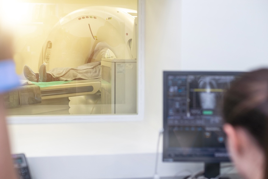
In their study, the researchers used an ultra-high field MRI scanner to observe a mouse model of PD. The experimental MRI machine, which generates a magnetic field of 9.4 Tesla—far stronger than the typical 3 Tesla used in medical settings—provided enhanced imaging, allowing for a detailed view of the mouse brain. The transgenic mice in the study carried elevated levels of alpha-synuclein, a protein implicated in PD. This protein accumulates and forms inclusions in the substantia nigra, the brain region responsible for dopamine production. The progressive degeneration of this area causes the motor symptoms seen in PD. The model used in this study is valuable because it mimics human alpha-synuclein accumulation and the resulting disease progression. Additionally, the behavior of these mice indicated an impaired sense of smell, and they likely experienced visual impairments as well. Unlike typical fMRI studies in animal models that focus on one sense, this research assessed both visual and olfactory sensory modalities, which is uncommon in fMRI experiments.
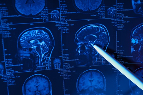
Functional MRI (fMRI) detects brain activation in response to stimuli, such as odors or visual input, by tracking changes in blood flow and oxygenation, which reflect neural activity. The researchers first compared fMRI results from mice with alpha-synuclein tangles to those of their healthy siblings. The control mice showed normal brain activity, while the PD mice exhibited significantly reduced activity. However, fMRI does not directly measure neural activity; instead, it captures a mix of neural and vascular effects, which complicates the interpretation. To separate these effects, the team used cerebral blood flow mapping to assess vascular changes and found that the PD mice had weaker vascular activity than the controls. Additionally, they measured neural activity by quantifying the C-FOS protein, which is released when neurons are activated. The post-mortem analysis revealed a more significant reduction in neuronal activity than in vascular flow in the PD mice. The researchers concluded that the changes observed in the fMRI scans were primarily driven by alterations in neuronal activity, though both vascular and neural factors were involved. This study marks a significant step forward in understanding early PD detection, and the approach may ultimately offer a way to diagnose PD years before clinical symptoms appear, allowing for earlier and more effective interventions.
“To our knowledge, this is the first observation of a combined visual and olfactory sensory aberration in the brain activity of PD rodent models in general and the alpha-synuclein model in particular”, the authors wrote in their paper published in the Journal of Cerebral Blood Flow and Metabolism. “This provides an opportunity for future studies to interrogate how sensory deficits progress along the disease, and perhaps lead to early imaging biomarkers [of the disease] (...).”
Related Links:
Champalimaud Foundation