AI Model Detects Diabetes Using Chest X-Rays
Posted on 04 Aug 2023
Present guidelines recommend screening individuals aged between 35 and 70 years who are overweight to obese, as indicated by their Body Mass Index (BMI), for type 2 diabetes. Nonetheless, numerous studies indicate that this approach fails to identify a significant number of cases, especially among racial and ethnic minorities for whom BMI is a less reliable indicator of diabetes risk. Undiagnosed diabetes patients are at a much higher risk of developing complications, including irreversible organ damage and even death. Now, a new artificial intelligence (AI) model has demonstrated that X-ray images taken during routine medical care can reveal signs of diabetes in individuals who do not meet the criteria for increased risk. This could aid doctors in detecting the disease earlier, preventing complications.
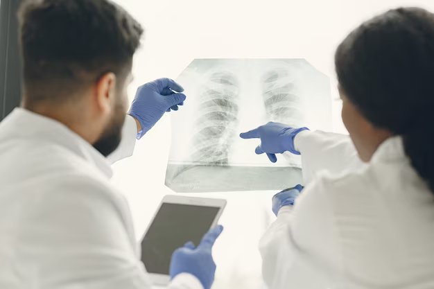
Using deep learning on images and electronic health record data, a multi-institutional team has developed a model that successfully flagged heightened diabetes risk in a retrospective analysis, often years before the disease was diagnosed. The AI model was trained on over 270,000 X-ray images from around 160,000 patients, with deep learning identifying the image features that best predicted a future diabetes diagnosis. As chest X-rays are not typically used for diabetes detection, the researchers utilized explainable AI techniques to understand the reasoning behind the model's predictions. The methods identified the location of fatty tissue as crucial in determining risk, which aligns with recent medical research linking visceral fat in the upper body and abdomen to type 2 diabetes, insulin resistance, hypertension, and other conditions.

Due to the groundbreaking nature of the approach and its remarkable results, the initial team enlisted researchers from Emory University (Atlanta, GA, USA) to externally validate the model. When applied to an independent group of nearly 10,000 patients, the model outperformed a basic model based on non-image clinical data in predicting diabetes risk. In certain cases, the chest X-ray flagged high diabetes risk up to three years before an eventual diagnosis. The model also provides a numerical risk score that could potentially aid clinicians in personalizing treatment plans for patients.
Annually, millions of chest X-rays are taken due to chest pain, difficulty breathing, injury, or as a pre-surgery procedure. While radiologists are not specifically looking for diabetes when examining these X-rays, such images become part of a patient's medical history and could be analyzed later for diabetes or other conditions. The researchers now plan to validate the model further and integrate it into electronic health record systems to alert physicians to conduct traditional diabetes screening for patients identified as high-risk based on X-ray findings. Their next focus will be to investigate the effectiveness of chest X-rays in diagnosing other conditions, such as vascular disease, congestive heart failure, and chronic obstructive pulmonary disease.
“Chest X-rays provide an ‘opportunistic’ alternative to universal diabetes testing,” said Judy Wawira Gichoya, MD, assistant professor of radiology and imaging sciences, and the lead researcher from Emory. “This is an exciting potential application of AI to pull out data from tests used for other reasons and positively impact patient care.”
Related Links:
Emory University


