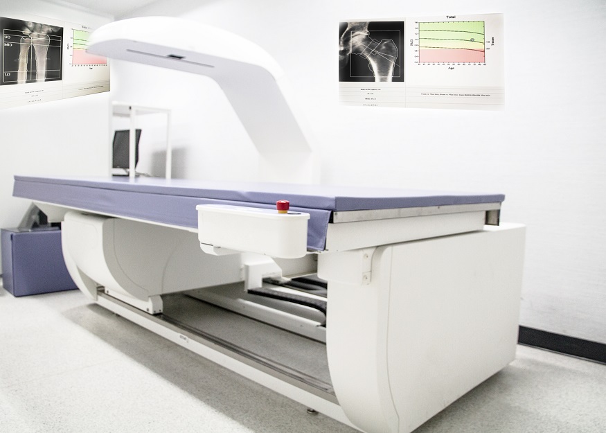
Bone Density Test Predicts Heart Attack Risk
Posted on 04 Aug 2023
A standard osteoporosis screening test, which measures bone density, can also detect an elevated risk for heart attacks due to the presence of calcium in the aorta. However, the interpretation of these images demands expertise and can be a time-consuming process. New research has now revealed that the use of machine learning to calculate this calcification test score can make the process faster and more efficient, eliminating the need for human evaluation of the scans and helping predict heart attack risk.
The task of scoring abdominal aortic calcification (AAC) from images produced by bone density machines is a painstaking process that requires meticulous training. Consequently, AAC scoring is not commonly carried out in clinical practice when these images are acquired. In a multi-institution research collaboration that included Harvard Medical School (Boston, MA, USA), scientists have developed, validated, and tested machine-learning algorithms for AAC assessment. This new tool, known as ML-AAC-24, was then evaluated in a real-world setting using a registry study of 8,565 older males and females. The researchers found that higher ML-AAC-24 scores were linked with considerably elevated cardiovascular disease risk and worse long-term prognosis.

“During DXA scans obtained for bone-mineral density testing, vascular calcification of the aorta can be seen and quantified,” said Naeha Sharif of Edith Cowan University. “This study developed a machine-learning algorithm to automatically determine the severity of the calcification that corresponds closely with the manual reading that is far more time-consuming to perform.”
“This development paves the way for use in routine clinical settings with little or no time to generate the useful calcification score that predicts heart attacks,” added Douglas Kiel, HMS professor of medicine and director of the Musculoskeletal Research Center at Hebrew SeniorLife.
Related Links:
Harvard Medical School


