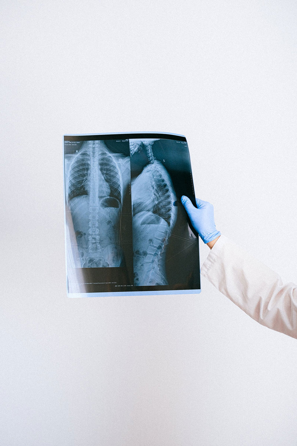
Simple X-Ray Method Can Diagnose Vertebral Compression and Prevent Spinal Fractures
Posted on 29 Jun 2022
Vertebral compression means that the spine is compressed, causing a fracture in one of the vertebrae. Vertebral compression fractures (VCFs) occur easily in people with osteoporosis and are very common among older people. However, the majority are unaware that these are causing their back pain – only one in three is diagnosed. Now, new research suggests that a simple X-ray method should be introduced as a routine procedure in healthcare so that more elderly patients can be diagnosed and given the most efficacious drugs.
A thesis by researchers at the University of Gothenburg (Gothenburg, Sweden) shows that VFA is of great clinical benefit, and the results suggest that the method should be introduced as a routine procedure in Swedish health care. Vertebral compressions do not necessarily result in distinct symptoms. Imaging is needed to detect these spinal fractures. If more people were diagnosed, many fractures, much suffering and heavy costs could be avoided.

In Sweden, efforts to prevent fractures in older people vary. In some regions, but not all, there are established “fracture liaison services,” as they are known in Sweden. These ensure that investigations of fractures are structured in a way that greatly reduces the risk of repeat fractures. When elderly patients have suffered fractures, their hip and lumbar spine bone density is examined with the dual-energy X-ray absorptiometry (DXA) method, to find out if they need treatment for osteoporosis. DXA can then also be used to get a side view of the chest and lumbar spine, with a method called vertebral fracture assessment (VFA), in which the height of the vertebrae is analyzed.
“The VFA method provides a very low radiation dose, and it’s fast, cheap, simple, and effective in finding vertebral compressions. It’s a valuable method for diagnosing relevant compressions, and significantly improves fracture risk assessment in older women,” said Lisa Johansson, doctoral student at Sahlgrenska Academy, University of Gothenburg, who authored the thesis.
Related Links:
University of Gothenburg


