Siemens’ New MRI Technology Offers Potential to Be Used for Lung Imaging Without X-Ray Radiation
Posted on 17 Dec 2021
A new MRI technology uses a lower magnetic field to open up new possibilities for imaging the lungs and patients with implanted devices and will potentially support new interventional procedures that could result in less radiation exposure.
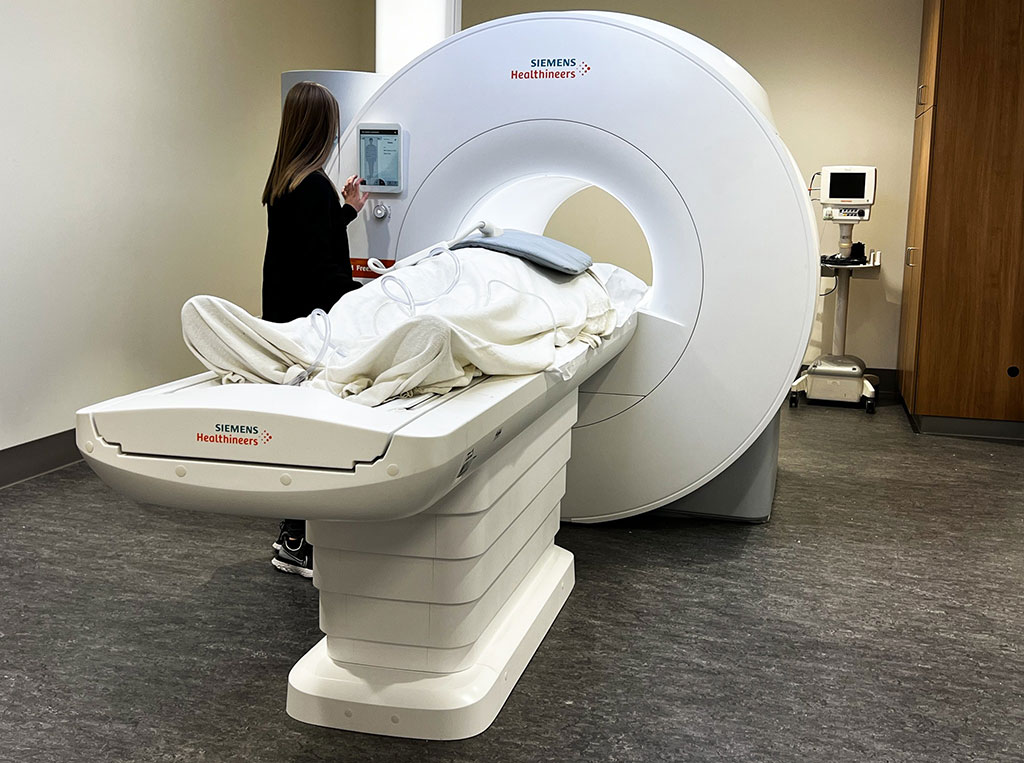
The new MRI technology, developed by Siemens Healthineers (Erlangen, Germany), in collaboration with researchers at The Ohio State University (Columbus, OH, USA), will expand imaging access for patients with implanted medical devices, severe obesity or claustrophobia. Siemens' recently FDA-approved 0.55T MAGENTOM Free.Max full body MRI for patient care features the largest MRI opening to date, 80 cm compared to the typical 60-70 cm, and a lower magnetic field strength that offers the potential for it to be used for lung imaging without X-ray radiation.

MRI uses a powerful magnetic field and radio waves to produce detailed images of a patient’s body to help diagnose conditions, plan treatments and determine the effectiveness of previous treatments. MRI is used predominantly to image the brain, spine and joints but can also be used to image the heart and blood vessels. Today’s clinical MRIs usually have magnetic field strengths of 1.5 or 3.0 Tesla, whereas the Free.Max is much lower at 0.55 Tesla.
“Many of our patients have pacemakers or defibrillators and while many of those devices are now safe for MR scanning, the metal in them can distort the magnetic field and corrupt the image quality. We were looking for ways to improve the quality of images in these patients, and lower magnetic field strength could offer an advantage. The problem with low field MRI is that there is less signal to work with, and we needed to find ways to boost that signal,” said Orlando Simonetti, research director of cardiovascular magnetic resonance, professor of Internal Medicine and Radiology and John W. Wolfe professor in cardiovascular research.
Ohio State then teamed up to develop techniques that could suppress noise, or interference in the images, and produce clearer images at lower field strength. They shared their ideas and techniques with Siemens, leading to the development of the 0.55T Free.Max scanner. The Ohio State University Wexner Medical Center is the first in the US to install the Free.Max as part of Ohio State’s cardiovascular imaging program. Ohio State researchers are optimistic that the new MRI technology can also be used to image the lungs, which typically is done with nuclear imaging or X-ray CT scans.
“This is an important advancement for patients with cystic fibrosis, pulmonary hypertension, heart failure, COVID-19 and any other disease where we’re trying to understand the source of shortness of breath and evaluate both the heart and lungs,” Simonetti said. “The air in the lungs cancels out the MRI signal at higher field strength; however, at lower field, there's potential to see lung tissue more clearly with the MRI.”
Related Links:
Siemens Healthineers
The Ohio State University


