New Algorithm for Rapid, Automated Diagnosis of COVID-19 from Chest CTs Overcomes RT-PCR Limitations
Posted on 25 Oct 2021
Scientists have developed a new algorithm for rapid, computerized diagnosis of COVID-19 that overcomes the limitations of reverse transcription polymerase chain reaction.
The new framework for accurate and interpretable automated analysis of chest CT scans was developed by researchers at the Daegu Gyeongbuk Institute of Science (DGIST; Daegu, South Korea). The current standard for diagnosis of COVID-19 through reverse transcription polymerase chain reaction (RT-PCR) is limited owing to its low sensitivity, high rate of false positives, and long testing times. This makes it difficult to identify infected patients quickly and provide them with treatment. Furthermore, there is a risk that patients will still spread the disease while waiting for the results of their diagnostic test.

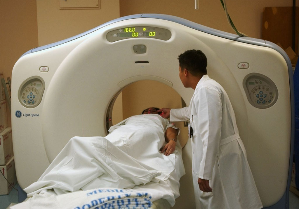
Chest CT scans have emerged as a quick and effective way to diagnose the disease, but they require radiologist expertise to interpret, and sometimes the scans look similar to other kinds of lung infections, like bacterial pneumonia. Now, a team of scientists have developed a technique for the automated and accurate interpretation of chest CT scans. To build their diagnostic framework, the research team used a Machine Learning technique called “Multiple Instance Learning” (MIL). In MIL, the machine learning algorithm is “trained” using sets, or “bags,” of multiple examples called “instances.” The MIL algorithm then uses these bags to learn to label individual examples or inputs.
The research team trained their new framework, called dual attention contrastive based MIL (DA-CMIL), to differentiate between COVID and bacterial pneumonia, and found that its performance was on par to other state-of-the-art automated image analysis methods. Moreover, the DA-CMIL algorithm can leverage limited or incomplete information to efficiently train its AI system. This research extends far beyond the COVID pandemic, laying the foundation for the development of more robust and cheap diagnostic systems, which will be of particular benefit to under-developed countries or countries with otherwise limited medical and human resources.
“Our study can be viewed from both a technical and clinical perspective. First, the algorithms introduced here can be extended to similar settings with other types of medical images. Second, the ‘dual attention,’ particularly the ‘spatial attention,’ used in the model improves the interpretability of the algorithm, which will help clinicians understand how automated solutions make decisions,” explained Prof. Sang Hyun Park and Philip Chikontwe from DGIST, who led the study.
Related Links:
Daegu Gyeongbuk Institute of Science (DGIST)


 Guided Devices.jpg)


