Novel CT System Provides Spatial Breast Imaging
By MedImaging International staff writers
Posted on 18 Oct 2021
An advanced cone beam computed tomography (CBCT) system dramatically improves the way clinicians can visualize, evaluate, and biopsy breast tissue. Posted on 18 Oct 2021
The Koning (Norcross, GA, USA) Koning Breast CT (KBCT) system is a new breast imaging platform that produces true three-dimensional (3D) images of the breast, without painful compression, in a simple, rapid 10-second procedure at the same radiation levels used during mammography. KBCT images are also less distorted than mammography, and are optimized so as to differentiate between normal and cancerous breast tissue. The KBCT acquires the entire volume of the breast, allowing radiologists to spatially scroll through the slices (up and down, left and right).

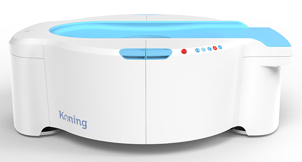
Image: The KBCT 3D breast imaging system (Photo courtesy of Koning)
The system consists of a horizontal patient table, the CBCT scanner mounted on a rotating assembly, an operator’s console, a reconstruction software engine, and a 3D visualization/temporary DICOM storage package. Data acquisition is achieved via a flat panel detector (FPD), an X-ray tube, and a 480 VAC high frequency generator. The tube/detector assembly rotates around the breast (which is located at the rotation axis), acquiring 300 projection images. Radiologists can also co-register data in multiple planes, similar to whole body CT imaging.
“Current breast imaging modalities have been lacking in their ability to confidently recognize breast cancer at earlier stages in its development,” said David Georges, President of Konig. “The Koning Breast CT can find lesions as small as 2mm and calcifications as small as 200 microns without breast compression.”
“Koning Breast CT is a true 3D dimensional image of the breast,’ said Professor Etta Pisano, MD, of Beth Israel Deaconess Medical Center (BIDMC; Boston, MA, USA). “The detector is surrounding the woman’s breast, and so the cancers have nowhere to hide in the breast. There’s no way for over lapping tissue to be in the way from every angle.”
During CBCT, the region of interest is centered in the field of view. A single 200 degree rotation acquires a volumetric data set which is used to produce a digital volume composed of 3D voxels of anatomical data that can then be manipulated and visualized. CBCT has only recently become practical with the introduction of large-area high-speed digital X-ray imagers, such as hydrogenated amorphous silicon (a-Si:H) based FPDs.
Related Links:
Koning
Beth Israel Deaconess Medical Center


