3D Data Used to Visualize Cardiac Conductive System
By MedImaging International staff writers
Posted on 14 Aug 2017
Researchers have discovered new details of how the conductive system of the human heart functions that could help cardiac surgeons repair hearts without damaging healthy tissue.Posted on 14 Aug 2017
The results of this pioneering study provide improved and more accurate computer models of the conductive system of the human heart, and the origins of the heartbeat, and could help improve clinicians’ understanding of atrial fibrillation and other common cardiac problems.

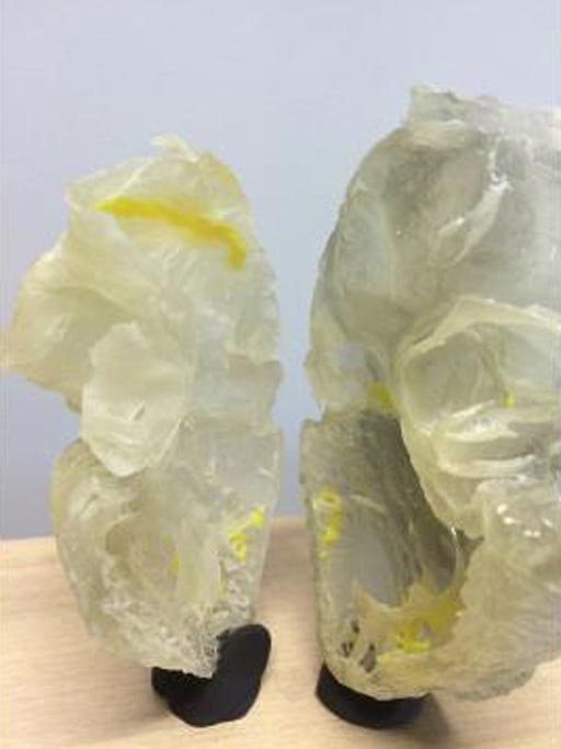
Image: A plastic 3D printed heart highlights the human cardiac conductive system (Photo courtesy of the University of Manchester).
The scientists from Liverpool John Moores University (LJMU; Liverpool, UK), The University of Manchester (Manchester, UK), Aarhus University (Aarhus, Denmark), and Newcastle University (Newcastle, UK) published the research findings online in the August 3, 2017, issue of the journal Nature, Scientific Reports.
The scientists soaked post-mortem samples of heart tissue in an iodine solution to enhance visualization of heart tissue in X-Ray images. They then used X-Ray scanners to make 3D images, some of which were so detailed that they showed the boundaries between individual heart cells, and the cellular layout in the tissue.
Professor Jonathan Jarvis, at the LJMU School of Sport and Exercise Sciences, said, "The 3D data makes it much easier to understand the complex relationships between the cardiac conduction system and the rest of the heart. We also use the data to make 3D printed models that are really useful in our discussions with heart doctors, other researchers and patients with heart problems. New strategies to repair or replace the aortic valve must therefore make sure that they do not damage or compress this precious tissue. In future work we will be able to see where the cardiac conduction system runs in hearts that have not formed properly. This will help the surgeons who repair such hearts to design operations that have the least risk of damaging the cardiac conduction system."
Related Links:
Liverpool John Moores University
University of Manchester
Aarhus University
Newcastle University


 Guided Devices.jpg)


