Diagnosis of Cardiac Sarcoidosis Improved by Diet Prior to Imaging
By MedImaging International staff writers
Posted on 28 Dec 2016
Researchers in the US have discovered that they can increase the certainty of diagnosing cardiac sarcoidosis, if from 72 hours until the diagnostic imaging exam the patients are subject to a high-fat, low-sugar diet.Posted on 28 Dec 2016
Sarcoidosis is caused by the inflammation of a number of organs, including the lungs and lymph nodes, and can eventually affect the heart as well, and cause death from pulmonary fibrosis. Cardiac Sarcoidosis is most often diagnosed using Positron Emission Tomography (PET) and Computed Tomography (CT) scans, and a radioactive tracer called fludeoxyglucose (FDG), but the results are sometimes difficult to interpret.

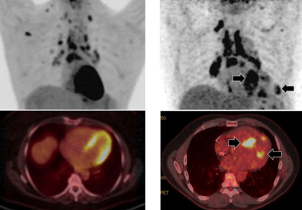
Image: The upper images shows PET scans, and the lower images combined PET/CT images of the heart. The images on the left show a patient on a 24-hour high-fat, low-sugar diet, while the right-hand images are those of a patient on a 72 hours high-fat, low sugar diet (Photo courtesy of UIC).
The researchers from the University of Illinois at Chicago (UIC; Chicago, Illinois, USA) published the results of their research online in the December 2016 issue of the journal Clinical Nuclear Medicine.
To reduce the amount of sugar taken up by healthy heart cells during the PET imaging, clinicians often give patients a high-fat, low-sugar diet for 24 hours before the scan. Healthy cells take up fat, while diseased cells take up the tracer and sugar, and are more easily detected in the scans. The UIC team then tried to use a 72-hour high-fat, low-sugar diet, before the scans, in 215 tests from 207 patients, in the study period between 2014 and 2015. The results showed that 42% of patients with the 24-hour diet had indeterminate results, compared to only 4% of patients with a 72-hour diet. The 72-hour diet enabled the researchers to conclude with a high degree of confidence whether the patients had cardiac sarcoidosis or not.
Corresponding author of the study, Dr. Yang Lu, assistant professor of radiology, UIC College of Medicine, said, "Up until now, diagnosis is made by expert opinion based on the results of several testing techniques including imaging. But what these scans show is often ambiguous, and so using them to definitively diagnose cardiac sarcoidosis is problematic. By following the diet for three days instead of one, the unpredictable but normal uptake of sugar by healthy heart cells was suppressed, so we could be more confident that areas that picked up the FDG had a very high likelihood of being affected by sarcoidosis, and this minimized the number of indeterminate findings where we can't make a diagnosis. The technique will also allow clinicians to better-follow their patients' progress under treatment for sarcoidosis."
Related Links:
University of Illinois at Chicago


