Study Suggests Teenage Obesity May Lead to Permanent Bone Loss
By Andrew Deutsch
Posted on 22 Nov 2016
Researchers have shown that obesity in adolescents affects bone density and could increase the risk of bone fractures in later life.Posted on 22 Nov 2016
Obesity is a major problem in many countries and is commonly associated with cardiovascular disease, and diabetes. The goal of the researchers is to try and find how obesity in adolescents affects bone structure.

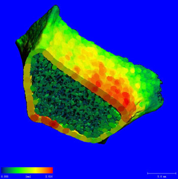
Image: A thickness map of the radius bone of the forearm, which was acquired using a SCANCO Medical Xtreme CT scanner (Photo courtesy of RSNA).
Twenty-three adolescents with a mean Body Mass Index (BMI) of 44 kg/m2, and with a mean age of 17 years took part in the study. The researchers used 3D High Resolution Peripheral Quantitative Computed Tomography (HR-pQCT) exams to measure bone microarchitecture, and mineral density in the arms and legs of the study participants. The scans enabled the researchers to study the structure of a bone in the forearm called the distal radius. In addition, the study participants underwent dual-energy X-Ray Absorptiometry (DXA) exams to quantify lean mass, and visceral fat mass. The research was presented at the annual Radiological Society of North America (RSNA2016) meeting.
The study results showed a positive association between BMI and cortical bone thickness and area, and cortical bone porosity. There was also a positive association between lean mass and trabecular density, bone volume, and integrity. The researchers concluded that a high amount of visceral fat combined with a low amount of muscle mass, was a risk factor for weakened bone structure in adolescents.
Lead author of the study, radiologist Miriam A. Bredella, MD, Massachusetts General Hospital (Boston. MA, USA), said, "While obesity was previously believed to be protective of bone health, recent studies have shown a higher incidence of forearm fractures in obese youths. Adolescence is the time where we accrue our peak bone mass, so bone loss during this time is a serious problem. We know from other chronic states that lead to bone loss in adolescence, such as anorexia nervosa, that increased fracture risk persists in adulthood, even after normalization of body weight. Therefore, it is important to address this problem early on. In addition, vitamin D, which is important for bone health, is soluble in adipose tissue and gets trapped within fat cells. The best way to prevent bone loss is a healthy diet that contains adequate amounts of calcium and vitamin D, along with sufficient exercise, as we have shown in our study that muscle mass is good for bone health."
Related Links:
Massachusetts General Hospital


