Details of Dinosaur Tail Revealed Using Spectral CT Technology
By MedImaging International staff writers
Posted on 03 Nov 2016
Paleontologists have made use of color spectral Computed Tomography (CT) images to obtain highly detailed images of the fossilized tail of a 66 million year old Tyrannosaurus Rex dinosaur.Posted on 03 Nov 2016
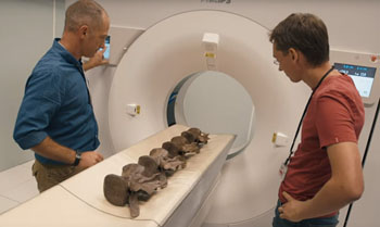
The scan of the tail vertebrae of the T. Rex Trix were scanned to compare spectral CT and conventional CT images, and to provide highly detailed images for the paleontologists.

Image: The researchers scanning the fossilized tail of a T. rex Trix dinosaur (Photo courtesy of Royal Philips).
The dinosaur skeleton was unearthed in Montana USA in 2013, and transported to the Naturalis museum in the Netherlands. The four tail vertebrae (C20 to C23) were scanned in color using the Royal Philips (Amsterdam, the Netherlands) IQon Spectral CT scanner in Best, the Netherlands. The Spectral IQon scanner has a unique dual-layer detector, and uses advanced algorithms for spectral reconstruction to provide detailed images of hard-to-scan objects.
Anne Schulp, dinosaur expert and paleontologist at the Naturalis museum, said, “We have already looked at some of the bones using a regular medical CT scanner, but we had a little problem. There is a deposit of pyrite – an iron rich mineral that blurs the complete picture because it absorbs the X-rays. So basically we required a different technology. The IQon filters out all the ‘noise’, as it were, thereby providing a really good insight into the bone structure and how it is built up. It's basically making the step from black and white movies to color. In this way, we are able to add depth to Trix’s medical records: we make the invisible visible. It's just really hard to describe the sensation of finding something, seeing something that no one else has ever seen.”
Related Links:
Royal Philips


