Researchers Improve Accuracy of Molecular Imaging for Cancer Patients
By MedImaging International staff writers
Posted on 22 Jun 2016
Researchers have unveiled a new method that enables clinicians to measure Lean Body Mass (LBM) for cancer patients, and improve staging of the cancer, and help monitor therapy.Posted on 22 Jun 2016
The new Computed Tomography (CT) procedure enables clinicians to obtain accurate LBM measurements and more precise molecular imaging results. The technique for measuring LBM can take into account changes in individual body composition.

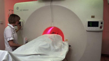
Image: A patient undergoing a positron emission tomography (PET) scan (Photo courtesy of Wellcome Images).
Researchers from the University of Alberta Edmonton (Alberta, Canada) presented the information in a scientific paper at the annual meeting of the Society of Nuclear Medicine and Molecular Imaging (SNMMI 2016) in San Diego, California, USA.
Clinicians routinely use positron emission tomography (PET) together with a radiotracer to find areas where there are abnormal increases in metabolic activity. The increase is measured using Standardized Uptake Values (SUV). The SUVs of radiotracer tumors are currently measured using the overall weight of a cancer patient, but this can change significantly during treatment, and can result in significant over or under-estimation of the LBM. The new CT method can measure patient-specific LBM much more accurately, and is called SULps.
Principal author of the study, Alexander McEwan, MD, University of Alberta Edmonton, said, "Patients with advanced cancer tend to lose muscle and may gain fat, and these changes in body composition can significantly modify PET results, independent of the actual metabolic activity of the tumor. Our study shows that CT-derived SULps is a more robust measurement for patients with advanced cancer undergoing PET imaging. If adopted, this simple change in imaging protocol could lead to significantly more effective care for cancer patients."
Related Links:
University of Alberta Edmonton


