Fate of Penumbra After Stroke Dependent on Blood Flow Restoration
By MedImaging International staff writers
Posted on 13 Jun 2016
A study investigating the fate of brain tissue that is at risk of dying after a stroke, has found that damage outcomes are association with collateral flow rather than time.Posted on 13 Jun 2016
The researchers found that treatment of the tissue at risk after a stroke, the penumbra, may need to be changed if the time of the stroke is unknown or treatment was delayed.

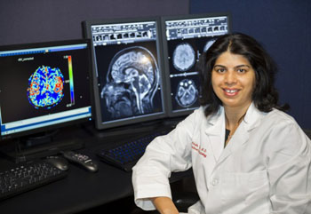
Image: Study leader Achala Vagal, MD, from the College of Medicine at the University of Cincinnati (Photo courtesy of University of Cincinnati).
The study that included 110 patients was led by an associate professor from the University of Cincinnati College of Medicine and radiologist at UC Health (UC; Cincinnati, OH, USA). The results were presented at the annual meeting of the American Society of Neuroradiology on May 25, 2016, in Washington, DC, USA.
The researchers did not find any significant correlation between salvaged penumbra tissue and time, but did find a correlation between the salvaged penumbra and the amount of collateral blood flow.
Study leader Achala Vagal, MD, said, “Using a large, multicenter stroke registry, we analyzed all untreated acute stroke patients who received baseline CT angiogram, an X-Ray that uses a dye and camera (fluoroscopy) to take pictures of the blood flow in an artery, and CT perfusion, to show which areas of the brain were getting blood, within 24 hours of the onset of stroke, and follow-up CT angiogram or MR angiogram within 48 hours. Baseline CT angiogram results were reviewed for artery blockages and rerouting of blood flow, and follow-up imaging was reviewed to determine if blood flow was restored.”
Related Links:
University of Cincinnati College of Medicine


