Functional Imaging Helps Test Spine Stability
By MedImaging International staff writers
Posted on 04 May 2016
A new functional imaging test called vertebral motion analysis (VMA) enables patients to demonstrate evidence of spinal instability.Posted on 04 May 2016
Developed by Ortho Kinematics (OKI; Austin, TX, USA) VMA testing involves standard surgical C-arms to capture moving “video x-ray” images using fluoroscopy. The images are uploaded to online servers, where they are processed to yield motion measurements that can be used to assess spinal instability. The cloud-based system also provides access to the images and test results via any internet-connected computer, tablet, or smartphone, providing an online tool that makes consulting with patients and interacting with insurance companies much easier.

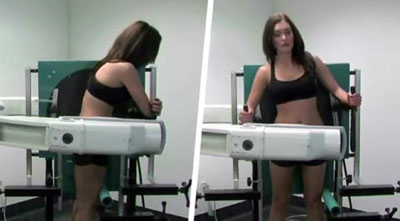
Image: Vertebral motion analysis testing (Photo courtesy of Ortho Kinematics).
The VMA lumbar spine quantitative fluoroscopy test takes about five minutes more than the standard 15-minute flex-ex test, but offers more robust data with less radiation exposure for both the patient and technologist. In peer-reviewed studies, VMA has been shown to be more accurate, repeatable, and five times more sensitive in detecting lumbar radiographic instability as compared to flex-ex testing, but just as specific. OKI has received U.S. Food and Drug Administration (FDA) and the European Community (CE) mark of approval for the VMA.
Since the 1940s, traditional flex-ex testing has been the standard of care for assessing instability based on spine motion analysis. Flex-ex testing requires physicians to measure spine motion from x-rays by hand. The results are highly variable, but because flex-ex data is so important, it is still ordered over five million times every year in the United States, more than spine computerized tomography (CT) and magnetic resonance imaging (MRI) imaging combined.
Related Links:
Ortho Kinematics


