Pioneering Technique for Imaging Biological Tissues Developed
By MedImaging International staff writers
Posted on 25 Aug 2015
Researchers have developed a novel X-ray imaging system that uses a compact X-ray source generated by ultra-short, high-power laser pulses, combined with phase-contrast X-ray tomography, to provide detailed 3-D imaging of tissues within organisms.Posted on 25 Aug 2015
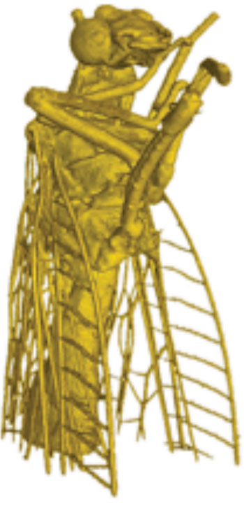
The new imaging system can be used to visualize very small structures, one-hundredth the diameter of a human hair, in 3-D, and can create images soft tissues. The technique uses phase-contrast X-ray refraction, unlike current radiographic techniques which are based on the absorption of X-rays. The physicists demonstrated the technique by producing an extremely detailed 3-D view of the cuticular structures of an insect.

Image: 3-D image of a fly using a new X-ray Imaging Technique (Photo courtesy of Nature Communications, and LMU).
The 3-D views were compiled by combining approximately 1,500 individual images, taken from different angles and assembling them into a 3-D data set.
The new technique could be used in the future to distinguish the difference between less-dense healthy tissue, and denser cancerous tissue, and could be used to detect early-stage tumors, less than 1 mm in diameter, before the can spread. The use of ultra-short X-ray pulses should also enable researchers to use the technique to freeze ultra-fast femtosecond processes, for example in molecules.
The new imaging system was developed by physicists at the Ludwig-Maximilians Universitat Munchen (LMU; Munich, Germany), the Max Planck Institute of Quantum Optics (MPQ; Garching bei Munchen, Germany), and the Technische Universität München (TUM; Munchen, Germany).
Related Links:
LMU
MPQ
TUM


