Computer Aided Detection of Tuberculosis in Chest Radiographs Reaches Maturity
By MedImaging International staff writers
Posted on 16 Feb 2015
Around nine million people get infected with tuberculosis (TB) every year. TB is still one of the most deadly diseases worldwide with over one million casualties annually, and early detection is critical to the success of treatment. Posted on 16 Feb 2015
Chest X-Rays have been used for detection of TB for many years, however it has always been difficult to interpret radiographs consistently. In addition, before digital chest radiography became available, the time to process radiographs was longer and the costs higher. Digital X-Ray systems are cheaper and faster to operate, and the images can immediately be sent to a central location for reading.
, and corresponding detection by the CAD system (right) (Photo courtesy of Radboud UMC).")
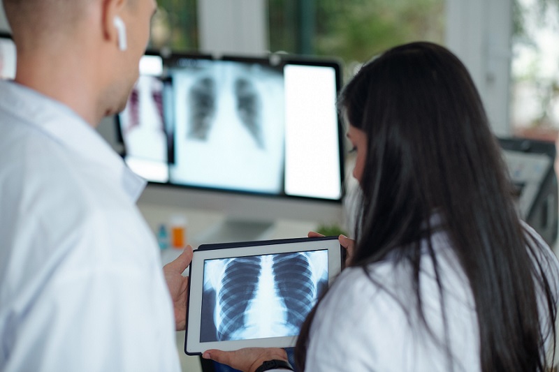
Image: Small lesion in the upper right lobe (left), and corresponding detection by the CAD system (right) (Photo courtesy of Radboud UMC).
.")
Image: Mobile Digital X-Ray Unit Used in the Gambia for TB Prevalence Survey (Photo courtesy of Radboud UMC).
To speed up detection and analysis of TB, and reduce costs, researchers at Delft Imaging Systems (Delft, The Netherlands), Radboud University Medical Center (UMC; Nijmegen, The Netherlands), and partners in the University of Cape Town Lung Institute (Cape Town, South Africa), and the NGO Zambart (Zambia) developed the CAD4TB software.
The CAD4TB software first scans the X-Ray image for correct posterior-anterior orientation during acquisition, and normalizes the image to make up for the differences between X-Ray machines.
CAD4TB consists of several separate systems which can automatically detect normal and abnormal structures in the lungs. The software is updated with machine learning techniques from thousands of existing radiographs. The data from the CAD4TB subsystems is compiled and the probability that an active TB infection exists in the patient are scored between 0–100.
The CAD4TB software is currently being evaluated in the Gambia, Tanzania, Pakistan, South Africa, and the Philippines, and the next release should have the CE marking (Conformité Européenne) certification.
Related Links:
Delft Imaging Systems
Radboud UMC
University of Cape Town Lung Institute

Gold Member
Solid State Kv/Dose Multi-Sensor
AGMS-DM+

Thyroid Shield
Standard Thyroid Shield

New
Wireless Handheld Ultrasound System
TE Air

New
X-Ray Detector
FDR-D-EVO III