Using Multiplanar Reconstructions to Predict Rupture of an Abdominal Aorta Aneurism
By MedImaging International staff writers
Posted on 02 Feb 2015
Rupture of an aneurism of the abdominal aorta is often fatal. Signs of frank rupture are clearly identifiable in Multidetector Computed Tomographic (MDCT) images, but an impending rupture is not as obvious. A rupture can lead to hemorrhage, limb or organ ischemia, or even death of the patient.Posted on 02 Feb 2015
To help radiologists identify aortic instability, researchers at the Russell H. Morgan Department of Radiology and Radiological Science, Johns Hopkins School of Medicine (Baltimore, MD, USA) published their research on pre- and post-operative indicators in the January-February 2015 issue of RadioGraphics journal.

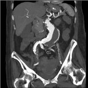
Image: Repeat contrast-enhanced multidetector CT including multiplanar reformations (Photo courtesy of Tonolini M, Department of Radiology, “Luigi Sacco\" University Hospital – Milan (Italy) and European Society of Radiology (ESR) EuroRAD).
Existing pre-operative Computed Tomography (CT) indicators can help radiologists recognize aortic instability and rupture. These indicators include a large initial size of the aorta, a rapid increase in the size of the aorta over a period of time, intramural hemorrhage (crescent sign), luminal expansion with lysis of thrombus, a penetrating atherosclerotic ulcer, periaortic hemorrhage, and contained rupture (draped aorta sign). Such changes need to be measured accurately and reliably with the help of Multiplanar Reconstructions (MPR), and semi-automated tools.
CT is routinely also used after an open or endovascular operation to keep track of graft complications. However the study shows that even when a surgical graft or endoluminal stent is in place aortic rupture can still occur.
Related Links:
Russell H. Morgan Department of Radiology and Radiological Science, Johns Hopkins School of Medicine


