Radiologist Recommendations for Chest CT Scanning Offer Important Findings
By MedImaging International staff writers
Posted on 13 Jan 2015
A significant proportion of patients who receive radiologist recommendations for undergoing chest computed tomography (CT) scanning to evaluate abnormal findings on outpatient chest X-rays have clinically relevant findings, including cancer, according to a new research. The investigators have reported that the findings demonstrate that radiologist recommendations for additional imaging (RAIs) after chest X-rays represent valuable contributions to patient care. Posted on 13 Jan 2015
The study was published online December 22, 2014, in the journal Radiology. RAIs, which have grown 200% since 1995, have drawn debate recently as the healthcare field transforms from volume-driven to value-based payment models. The scrutiny makes it increasingly important for the radiology community to confirm the clinical impact of its studies, according to study author Tarik K. Alkasab, MD, PhD, from Massachusetts General Hospital (MGH) and Harvard Medical School (both based in Boston, MA, USA). “There has been a great deal of research on how radiologists recommend an imaging exam, but little on what comes out of the exams that they recommend,” Dr. Alkasab said. “Prior studies were very broad, so in our study we tried to focus on a specific clinical scenario.”
Dr. Alkasab and colleagues examined chest X-rays, one of the most extensively performed outpatient diagnostic imaging studies in the United States. As many as 50% of all RAIs identified in thoracic diagnostic exams are the result of performing chest X-rays. The researchers searched through more than 29,000 reports of outpatient chest X-rays performed at a large academic center over one year to identify studies that included a recommendation for a chest CT. They found that radiologists interpreting outpatient chest X-rays made recommendations for CT in 4.5% of cases—a result in line with existing research. Increasing patient age and positive smoking history were associated with an increased likelihood of a chest CT recommendation.
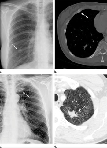
When the researchers examined the chest CT scans obtained within one year of the index chest X-ray, they discovered that 41.4% detected a corresponding abnormality requiring treatment or further diagnostic workup. One in every 13 generated a corresponding abnormality representing a newly-diagnosed, biopsy-validated malignancy.
“In this era of concern about radiation dose risk, these findings suggest that the extremely low predicted risk of radiation-induced cancer associated with a chest CT is orders of magnitude less than the potential clinical benefits,” said study coauthor H. Benjamin Harvey, MD, JD, from Massachusetts General Hospital and Harvard Medical School. “If ordering physicians see a recommendation for chest CT, they need to ensure that the patient gets the recommended imaging.”
More than one-third of patients in the study group who were recommended for a follow-up chest CT did not receive the exam within one year—an oversight that could result in missed or delayed diagnoses, the researchers said. “More research is needed to understand the possible reasons for the less-than-optimal adherence to RAIs after chest X-ray,” Dr. Harvey said. “One thing we’re looking at is how the recommendation language affects recommendation adherence.”
The researchers hope that their study helps enhance awareness of the importance of follow-up CT scanning. “These results show that radiologists should be confident their recommendations are adding value and protecting patients,” Dr. Alkasab concluded.
Related Links:
Massachusetts General Hospital


