X-Ray Imaging Components Optimized for Flexibility and Fast Patient Throughput
By MedImaging International staff writers
Posted on 17 Dec 2014
X-ray imaging components have been designed for high-speed, wireless, and advanced digital imaging systems.Posted on 17 Dec 2014
The components were developed by Varian Medical Systems (Palo Alto, CA, USA) and presented at the 2014 annual meeting of the Radiological Society of North America (RSNA) in Chicago (IL, USA), in December 2014. “As the industry adopts digital imaging technology, we’re able to help our customers with comprehensive solutions that combine X-ray tubes and digital detectors, and image processing software,” said Carl LaCasce, vice president sales and marketing for Varian imaging components. “Our components are optimized for digital imaging systems that support high patient throughput, flexibility, and usability. Our latest components and solutions are already designed into new systems for digital mammography, mobile C-arm and CT imaging.”
At the RSNA meeting, Varian presented X-ray tubes, PaxScan wireless digital image detectors, and Nexus image-processing software and workstations. Nexus software and workstations process data from mixed inputs, including older systems, radiofrequency (RF), and digital radiography (DR) flat-panel image detectors. “This advanced image processing software enables equipment manufacturers to add digital dynamic or radiographic productivity to their product portfolios, reducing their time to market and enhancing their competitiveness,” said Mr. LaCasce.
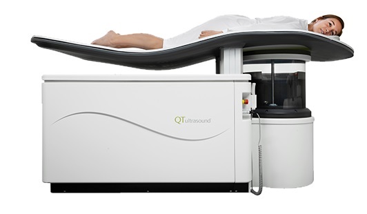
Varian’s mammography components are designed for systems that offer greater patient throughput and high power approaches, including tomosynthesis. “Our X-ray tubes produce images with high contrast at low doses in advanced imaging applications,” stated Mr. LaCasce. Varian’s newer digital tubes feature fan or liquid cooled housings with high heat dissipation rates. All can be used with standard three inch inserts. Varian also offers a compact full-field digital detector with a narrow edge that helps radiologists acquire images close to the chest wall.
Varian’s range of original equipment manufacturer (OEM) and replacement CT tubes utilize glass or ceramic and both bi-polar and anode end grounded (AEG) designs. The AEG tubes are designed for use at high power, with higher heat load and a greater ‘G’ force capacity, making them not only powerful but also durable. In addition, they offer reduced dose delivery outside the focal area.
PaxScan wireless digital image detectors enable equipment manufacturers to construct digital imaging systems that provide excellent image quality at high levels of throughput. Significant workflow efficiencies can be exploited during system design, to enhance the patient experience.
“Our goal is to reduce the time it takes for imaging with technical advances that improve the performance of the components that are at the heart of imaging systems,” said Mr. LaCasce. “With this technology, X-ray imaging can be more cost efficient and accessible for patients around the world.”
Related Links
Varian Medical Systems

Gold Member
Solid State Kv/Dose Multi-Sensor
AGMS-DM+

New
Pre-Op Planning Solution
Sectra 3D Trauma

New
Wireless Handheld Ultrasound System
TE Air

Brachytherapy Planning System
Oncentra Brachy