Personalized Intraoperative Radiation Therapy During Cancer Surgery
By MedImaging International staff writers
Posted on 09 Sep 2014
A New York City Medical Center is finding new ways to employ personalized, internal radiation delivered in the operating room right after a cancer tumor is removed. Intraoperative radiotherapy (IORT) represents an effort to reduce the risk of a recurrence, lessen the duration of standard postoperative external radiation, and reduce the risk to healthy tissue associated with external radiation. Posted on 09 Sep 2014
NewYork-Presbyterian Hospital Medical Center (New York, NY, USA), in 2012, became the first hospital in New York City to offer IORT to women with certain breast cancers. In this therapy, a spherical applicator is used to deliver a single, even dose of radiation to the inside surface of a rounded cavity after a lumpectomy.

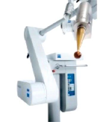
Image: The Zeiss Intrabeam mobile radiation oncology platform (Photo courtesy of Carl Zeiss).
Now, physicians at NewYork-Presbyterian/Columbia are expanding these innovating efforts by offering IORT for other types of cancer in the abdomen and pelvis. Unlike that in the breast, the tumor bed in the abdomen and pelvis may not be as precisely defined after surgery, and several sites at risk for recurrence may need to be treated.
Earlier in 2014, in the hospital’s first case of using IORT for a cancer other than breast cancer, a woman with recurrent colon cancer in the pelvic cavity needed to have treatment to separate areas of her body. The surgeon, Ravi Kiran, MD, a professor of surgery (in epidemiology) and chief of colorectal surgery at NewYork-Presbyterian/Columbia, removed the tumor, but could not cut too close to major blood vessels and other organs. Because of the fundamental limitations of surgery, and because the patient had already received a high lifetime cumulative dose of radiation therapy in earlier treatments, Dr. Kiran and Clifford Chao, MD, a professor of radiation oncology and chair of radiation oncology at NewYork-Presbyterian/Columbia, decided to use IORT to “mop up” any reamining tumor cells. Dr. Chao used a flat radiotherapy applicator to deliver radiation to areas close to blood vessels along the pelvic wall and a spherical applicator to treat a region lower in the pelvic cavity. “We also used a protective wrap, or draping, made of material that shields organs like the bowel or blood vessels from scatter radiation,” Dr. Chao said.
In a second procedure, which involved a 23-year-old woman with a bile duct tumor, two differently shaped applicators were used to deliver IORT to the retroperitoneal tissue after removal of the tumor and nearby lymph nodes. The surgery, performed by Tomoaki Kato, MD, a professor of surgical oncology and chief of abdominal organ transplantation at NewYork-Presbyterian/Columbia, was the first case in the United States to use Zeiss Intrabeam flat applicators after removal of a bile duct tumor. The flat applicator was used to deliver radiation to the surface where the retroperitoneal aortic lymph node had been. A sphere-shaped applicator was inserted into the right lobe of the liver near the hepatic artery, where the soft organ could completely surround the tiny globe and absorb a 20–30 minute, low-dose IORT session.
“Because of the way the tumor needs to be removed, or because the spaces between a tumor and large vessels and nerves are too small, microscopic lesions are more likely to be attached to the surface of blood vessels and nerves. IORT allows us to treat those areas and lower the risk of recurrence,” said Dr. Chao, who performed the radiation therapy procedure and is leading the hospital’s IORT efforts.
In a third patient, Jason Wright, MD, a professor of Women’s Health (in obstetrics and gynecology) and chief of gynecologic oncology at NewYork-Presbyterian/Columbia, used a similar approach to treat a gynecologic cancer, working with Dr. Chao. In specific breast cancer patients, IORT has eliminated an additional six to seven weeks of radiotherapy, and according to a 10-year randomized trial published in 2010, provided the same results as traditional full-breast radiation. Dr. Chao is hopeful that similar benefits will be seen in other types of cancer cases, and he sees this as another step toward customized cancer care.
“The possibilities are encouraging,” said Dr. Chao. “We could see patients ahead of time and then work with the surgeon to develop a personalized radiation treatment for the specific tumor. When you open up the abdomen to remove a tumor from the liver, bowel, or pancreas, the terrain of the surgical bed is a more open, uneven surface. So we need radiotherapy applicators that suit the specific anatomical terrain. In some areas of the body, the applicator could be a half sphere, an irregular shape for uneven surfaces, or a tiny device that fits into a small space where we have anatomic challenges. We can devise personalized therapy based on a patient’s specific anatomy.”
Dr. Chao is currently working with engineers and physicists from NewYork Presbyterian/Columbia and NewYork-Presbyterian/Weill Cornell Medical Center to design and develop applicators for colorectal, head and neck, lung, and gynecologic cancers.
Zeiss Intrabeam, developed by Carl Zeiss (Oberkochen, Germany), is a mobile radiation oncology platform that offers additional treatment options for a wide range of cancer types, through use of its US Food and Drug Administration (FDA)-cleared spherical, needle, flat, and surface applicators. Because of the low X-ray energy, Intrabeam does not require any complicated radiation shielding measures and is suitable even for mobile use.
Related Links:
NewYork-Presbyterian Hospital Medical Center
Carl Zeiss


