Self-Assembling Nanoparticles Could Improve Cancer Diagnosis
By MedImaging International staff writers
Posted on 21 Jul 2014
Innovative nanoparticles boost the effectiveness of magnetic resonance imaging (MRI) scanning by specifically seeking out receptors that are found in cancerous cells.Posted on 21 Jul 2014
Developed by researchers at Imperial College London (United Kingdom), the iron oxide nanoparticles (IONPs) were synthesized to selectively undergo copper-free click conjugation upon sensing of matrix metalloproteinase (MMP) enzymes, thereby leading to a self-assembled superparamagnetic-nanocluster network, with T2 signal enhancement properties. The IONPs were synthesized and characterized to tether to C-X-C chemokine receptor type 4 (CXCR4, also known as fusin) -targeted peptide ligands.

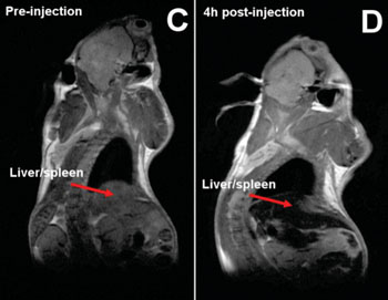
Image: Whole body images of a mouse before and after nanoparticles injections. Signal loss in the liver and the spleen due to the accumulation of iron from the nanoparticles is indicated by the red arrows. (Photo courtesy of Imperial College London).
The nanoparticle is coated bio-orthogonal azide and alkyne surfaces masked by polyethylene glycol (PEG) layers. When it finds a tumor it begins to interact with the cancerous cells, resulting in the stripping off of the PEG coating, causing the nanoparticle to self-assemble into a much larger particle so that it is more visible on the scan. The researchers tested the IONPs in vitro in rats, resulting in T2 signal enhancements of around 160%. Simultaneous systemic administration of the bio-orthogonal IONPs in tumor-bearing mice demonstrated the signal-enhancing ability of the self-assembling nanomaterials. The study was published online on July 15, 2014, in Angewandte Chemie.
“By improving the sensitivity of an MRI examination, our aim is to help doctors spot something that might be cancerous much more quickly, “ said senior author Prof. Nicholas Long, PhD, of the ICL department of chemistry at Imperial College London. “The results show real promise for improving cancer diagnosis. This would enable patients to receive effective treatment sooner, which would hopefully improve survival rates from cancer.”
“We're now looking at fine tuning the size of the final nanoparticle so that it is even smaller but still gives an enhanced MRI image,” added Juan Gallo, MD, of the department of surgery and cancer. “If it is too small the body will just secrete it out before imaging, but too big and it could be harmful to the body. Getting it just right is really important before moving to a human trial.”
Related Links:
Imperial College London


