Portable 3D Ultrasound System Enables Reproducible Breast Cancer Monitoring
Posted on 06 Jul 2026
Breast cancer can develop between annual mammograms, and these interval cancers account for 20% to 30% of cases and tend to be more aggressive. The challenge is pronounced in people with dense breast tissue. More frequent, operator‑independent imaging could also help with longitudinal monitoring after treatment. A new study shows a portable, real‑time three‑dimensional ultrasound system that improves resolution and standardizes probe placement.
Researchers at the Massachusetts Institute of Technology (MIT) developed the 3D portable ultrasound for real-time examination (PURE) system, a compact probe-and-module platform designed for operator‑independent breast imaging. The setup pairs a small ultrasound probe with an acquisition and processing module that is slightly larger than a smartphone. A computer‑vision interface guides users to the same anatomical location across sessions, enabling reproducible scans suitable for longitudinal follow‑up.
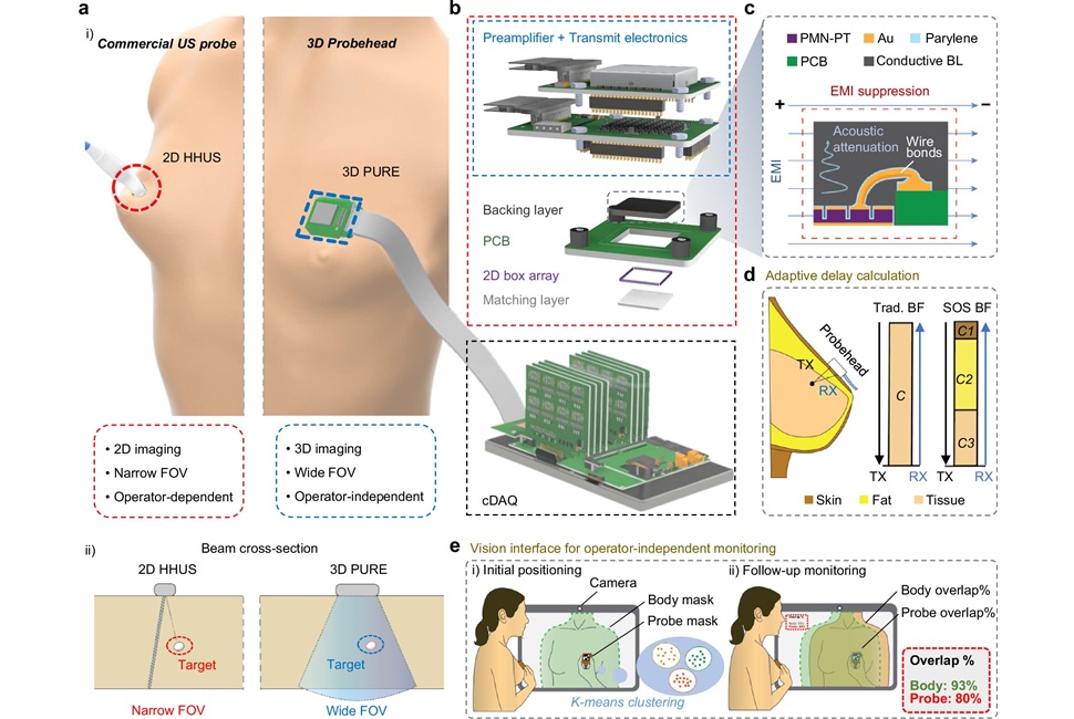
The technology combines hardware and software improvements to enhance image quality. Its ultrasound transducer incorporates a backing layer that contains and focuses acoustic energy, broadens the usable frequency range, and reduces both acoustical and electrical noise for sharper imaging. During reconstruction, an adaptive beamforming algorithm compensates for tissue-dependent speed-of-sound differences, such as those in skin and fat, producing up to a 10% increase in image resolution. The system can also generate a 3D rendering of the entire breast from two or three scan positions with live visualization.
Study details published in Nature Communications on July 1, 2026, describe assessments of usability and target detection. Ten volunteers without ultrasound expertise used the system to locate small microtargets embedded in a tissue phantom and achieved a much higher detection success rate than with a conventional probe. In a separate trial with seven people, users accurately repositioned the probe to the prescribed location on repeated scans, supporting consistent placement for longitudinal monitoring.
According to the authors, the platform could facilitate earlier diagnosis and long‑term surveillance after treatment in clinical settings or at home. The team also aims to adapt the interface for mobile devices and notes potential soft‑tissue applications beyond breast imaging, including ovarian cancer, endometriosis, and fetal monitoring.
“At each time interval, the computer interface guides you to position the device in exactly the same location, which is important for the longitudinal monitoring of a given tissue. It’s very intuitive and quite easy to use,” said Canan Dagdeviren, associate professor of media arts and sciences at MIT and the senior author of the study.
“What we are trying to do is predict the speed of sound properties of the tissue you’re imaging and then use that to reconstruct the image more accurately. We see up to a 10% improvement in the resolution just by applying this technique,” said Shrihari Viswanath, MIT graduate student.
Related Links
MIT