Pigeons Could Advance Pathology Image Analysis Tools
By MedImaging International staff writers
Posted on 09 Dec 2015
Pigeons can distinguish between healthy and cancerous tissue in X-rays and microscope slides with an accuracy rate of up to 99%, according to a new study.Posted on 09 Dec 2015
Researchers at the University of California Davis (UCD; USA) conducted a series of three experiments in Pigeons (Columba livia), to examine if their visual system properties could help them serve as surrogate observers of medical images. Research over the past 50 years has shown that pigeons can be prodigious discriminators of complex visual stimuli, and can discriminate among misshapen pharmaceutical capsules; recognize letters of the alphabet; categorize objects such as cats, flowers, cars, or chairs; identify emotional expressions and features of human faces; and even identify paintings styles.

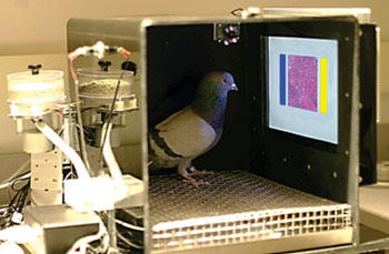
Image: The pigeons’ training environment, with a food pellet dispenser and a touch-sensitive screen with yellow and blue selection buttons (Photo courtesy of UCD).
During the first experiment, eight pigeons were presented with 144 breast tissue images, at various levels of magnification and with and without color. The birds were then trained to peck a blue or yellow button on either side of each image to indicate whether it was cancerous or healthy; if they chose correctly, they were rewarded with food. But if they chose incorrectly, they were presented with the image again and again until they correctly identified it. After 15 days of training, the birds’ accuracy rate had risen from 50% to 85%.
The pigeons were then presented with new images to rule out memorization as a possible cause for their success. They subsequently correctly identified familiar and novel images 87% and 85% of the time, respectively. When the researchers combined the birds’ responses, a method they called “flock sourcing,” rather than scoring them individually, they found that accuracy rates increased to 99%. In the second experiment, four new birds were tested to see if they could identify microcalcifications in breast tissue. Within 14 days, their accuracy rate rose from 50% to over 85%.
The final experiment had another four birds attempt to identify masses in mammograms. While the pigeons were able to identify masses in images they had already seen with some success, with two pigeons reaching 80% correct classification and two reaching 60%, they could not identify masses as benign or malignant in new images. According to the study, this can also be very challenging for humans; a panel of radiologists previously tested reached an 80% accuracy rate when viewing the same images. The study was published on November 18, 2015, in PLOS One.
“Pathologists and radiologists spend years acquiring and refining their medically essential visual skills, so it is of considerable interest to understand how this process actually unfolds, and what image features and properties are critical for accurate diagnostic performance,” said lead author professor of pathology and laboratory medicine Richard Levenson, MD, and colleagues. “The birds' successes and difficulties suggest that pigeons are well-suited to help us better understand human medical image perception, and may also prove useful in performance assessment and development of medical imaging hardware, image processing, and image analysis tools.”
Pigeons have are able to generalize their discrimination performance to novel objects or scenes, successfully differentiating among complex and varied visual stimuli. Color, size, shape, texture, and configural cues all seem to participate in the discrimination process. Importantly, the anatomical neural pathways involved, including basal ganglia and cortical-striatal synapses, appear to be functionally equivalent to those in humans.
Related Links:
University of California Davis


