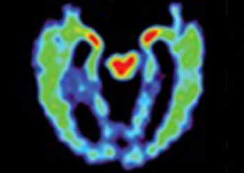
PET Scan Could Help Diagnose Head Trauma Damage
By MedImaging International staff writers
Posted on 28 Sep 2016
Proteins are deposited in a characteristic pattern in the brains of athletes suffering from in chronic traumatic encephalopathy (CTE), according to a new study.Posted on 28 Sep 2016
Researchers at the University of California, Los Angeles (UCLA; USA), NorthShore University Health System (NSUHS; Evanston, IL, USA), and other institutions conducted a study in 14 retired national football league (NFL) football players, all of whom sustained at least one concussion during their career. Study participants underwent a positron emission tomography (PET) scan using a chemical marker called FDDNP, which binds to deposits of neurofibrillary tau tangles and amyloid beta plaques. The PET scans were also compared to those of 28 men and women with healthy brains, and 24 men and women with Alzheimer’s disease (AD), all of similar ages.

Image: A PET scan of a brain with suspected CTE; red and yellow indicate abnormal brain protein (Photo courtesy of UCLA).
The results revealed that the retired football players showed tau deposit patterns consistent with those previously observed in autopsy studies of people with CTE, which indicated that brainstem white matter tracts undergo early axonal damage and cumulative axonal injuries along subcortical, limbic, and cortical brain circuitries supporting mood, emotions, and behavior. The deposition pattern was different from the progressive pattern of neuropathology in AD, which typically begins in the medial temporal lobe and progresses along the cortical default mode network, with minimal involvement of subcortical structures.
In addition, the areas in the brain where the patterns occurred were also consistent with the types of symptoms experienced by some of the study participants. When compared with healthy people, the former athletes had higher levels of FDDNP in the amygdala and subcortical regions of the brain, while people with AD had higher levels of FDDNP in areas of the cerebral cortex. Athletes who suffered more concussions also had higher FDDNP levels. The study was published in the June 2016 issue of Proceedings of the National Academy of Science (PNAS).
“We found that the imaging pattern in people with suspected CTE differs significantly from healthy volunteers and those with Alzheimer’s dementia,” said study author Julian Bailes, MD, director of the Brain Injury Research Institute at NSUHS. “These results suggest that this brain scan may also be helpful as a test to differentiate trauma-related cognitive issues from those caused by Alzheimer’s disease.”
CTE is thought to cause memory loss, confusion, progressive dementia, depression, suicidal behavior, personality changes, and abnormal gaits and tremor. Currently, it can only be diagnosed definitively following autopsy. To help identify the disease, doctors look for an accumulation of tau in the regions of the brain that control mood, cognition, and motor function. Tau is also one of the abnormal protein deposits found in the brains of people with AD, although in a distribution pattern that is different from that found in CTE.
Related Links:
University of California, Los Angeles
NorthShore University Health System


.jpg)
