Minimally Invasive Procedure Reduces Knee Osteoarthritis Pain
Posted on 19 Jun 2026
Knee osteoarthritis causes chronic inflammation, stiffness, and pain that impair mobility and daily function. Many patients exhaust injections and medication without durable benefit yet are not ready or eligible for joint replacement, creating a persistent treatment gap. This gap drives disability and ongoing utilization of health services. Researchers have now evaluated a minimally invasive vascular procedure designed to reduce pain and improve function without surgery.
In a prospective single-center study published in Radiology, researchers, including interventional radiologists at Charité-Universitätsmedizin Berlin, assessed genicular artery embolization (GAE) using rapidly resorbable gelatin-based microspheres for osteoarthritis-related knee pain. The approach was studied in adults who had not responded to at least three months of conservative therapy. Investigators aimed to establish safety and durability of symptom relief over 12 months.
. (A) Periinterventional image of the popliteal artery. Yellow arrow indicates the descending genicular artery, and blue arrow indicates the medial inferior genicular artery. (B, C) Selective preinterventional images show hypervascularity of two branches of the genicular artery: the (B) descending genicular artery (arrow) and the (C) medial inferior genicular artery (arrow). (D, E) Selective postinterventional images obtained after embolization with rapidly resorbable gelatin-based microspheres (Photo courtesy of RSNA)")
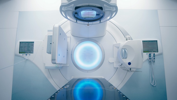
GAE targets abnormal hypervascularity that forms around the osteoarthritic joint. Under fluoroscopic guidance, a catheter is advanced to affected genicular artery branches, where size-calibrated microspheres are injected to occlude pathological vessels. The resorbable particles dissolve within hours, a strategy intended to combine the advantages of temporary and permanent embolic materials while limiting their drawbacks.
The study enrolled 194 participants (median age 69; median body mass index 28.4) and encompassed 239 technically successful procedures performed between July and November 2024. Twenty-three percent underwent staged bilateral treatment within four weeks. There were no moderate or severe adverse events, and only 6.7% experienced mild, self-limited reactions. Follow-up occurred at six weeks and at three, six, and 12 months, with a six-month in-person assessment by an orthopedic surgeon.
Median Numeric Rating Scale pain scores decreased from 7 at baseline to 4 by six weeks and to 3 at both six and 12 months. Knee Injury and Osteoarthritis Outcome Score sub-scores improved across symptoms, pain, daily activities, sports and recreation, and quality of life, with changes meeting accepted minimum clinically important differences. At 12 months, 80% of participants exceeded the clinically meaningful pain reduction threshold.
"Our study demonstrates that GAE using rapidly resorbable gelatin-based microspheres is a safe, minimally invasive therapy that provides meaningful pain relief and functional improvement in participants with osteoarthritis-related knee symptoms for at least 12 months," said Florian Nima Fleckenstein, M.D., deputy head of interventional radiology at Campus Mitte, Charité-Universitätsmedizin Berlin.
Related Links
Charité-Universitätsmedizin Berlin
RSNA