PET Technique Detects Bacterial Infections
By MedImaging International staff writers
Posted on 16 Oct 2017
A new non-invasive PET imaging technique has been developed that can detect bacterial infections, and monitor antibiotic therapy.Posted on 16 Oct 2017
Bacteria are developing antibiotic resistance by mutating, and this has led government healthcare authorities to call for the development of new diagnostic techniques that can detect and help manage infectious diseases.

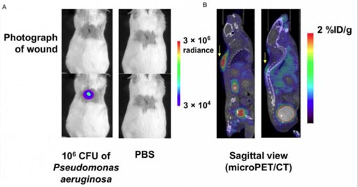
Image: Mice with a P. aeruginosa-infected wound (left) and control mice (right), and sagittal slices from a micro PET/CT scan one hour after administration of the new tracer (Photo courtesy of Sam Gambhir, MD, PhD, Stanford University).
The scientists from the Stanford University (Stanford, CA, USA) published their research, part of a pre-clinical study, in the October 1, 2017, issue of The Journal of Nuclear Medicine.
The researchers developed a new Positron Emission Tomography (PET) tracer called 6"-18F-fluoromaltotriose that is derived from maltose, labeled with radioactive fluorine-18 (18F), and evaluated it with a number of clinically-relevant bacterial strains, and a micro-PET/CT scanner.
The results show that both gram-negative, and gram-positive bacteria took up the new tracer, and that the tracer could be used to diagnose most bacterial infections, and could change the clinical management of bacterial infectious diseases in patients.
Sanjiv Sam Gambhir, MD, PhD, Stanford University, said, "The hope is that in the future when someone has a potential infection, this approach of injecting the patient with fluoromaltotriose and imaging them in a PET scanner will allow localization of the signal and, therefore, the bacteria. And then, as one treats them, one can verify that the treatment is actually working – so that if it's not working, one can quickly change to a different treatment (for example, a different antibiotic). These kinds of findings are very important for patients, because they will very likely lead to entirely new ways to manage patients with bacterial infections, no matter where those infections might be hiding in the body."
Related Links:
Stanford University


