New Hybrid PET/MRI System Developed for Enhanced Breast Cancer Imaging
By MedImaging International staff writers
Posted on 02 Feb 2016
A novel imaging technique is being developed that can improve breast cancer detection and characterization, and help clinicians evaluate the response of the cancer to treatment.Posted on 02 Feb 2016
The 4-year project, named Digital Hybrid Breast PET/MRI for Enhanced Diagnosis of Breast Cancer (HYPMED), began on January 1, 2016, and is being developed under a new cross-European project coordinated by the European Institute for Biomedical Imaging Research (EIBIR; Vienna, Austria). Other HYPMED project partners include the University Hospital Aachen (UKA; Aachen, Germany), and four other research institutes in Germany and The Netherlands. Four industry partners in Germany, France, and the Netherlands, including Royal Philips (Amsterdam, The Netherlands) are also taking part.

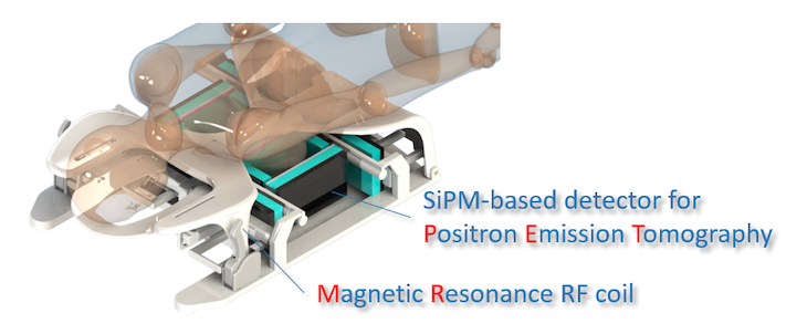
Image: The SiPM detector (Photo courtesy of Volkmar Schultz, Aachen University).
Breast cancer is still the most common female cancer, and a major cause of cancer death in women. The stage at which breast cancer is detected is at the moment the most important factor indicating survival of the patient, and there is a pressing need to improve early diagnosis of the disease.
The HYPMED project will develop a hybrid Magnetic Resonance Imaging (MRI) and Positron Emission Tomography (PET) imaging system intended to improve diagnosis of breast cancer, and provide personalized control of therapy results. The consortium intends to develop a Radio Frequency (RF) coil that can used in any standard clinical MR scanner, transforming the scanner into a high-resolution hybrid PET/MRI system that can identify extremely small breast cancer foci, better characterize the cancer, and improve treatment response. The radiation dose of the new system is expected to be minimal, comparable to a regular digital mammogram. The consortium also plans to use the HYPMED approach in the future for prostate cancer detection, hybrid cardiac imaging, and other diseases.
Scientific Coordinator of the project, Professor Christiane Kuhl, University Hospital Aachen, said, “The HYPMED project combines visionary clinical expertise with excellence in physical and engineering sciences and the developed technology will greatly help us to choose an appropriate treatment that is exactly right for a given cancer in a given woman.”
Related Links:
EIBIR
UKA
Royal Philips


