Single PET Scan Could Replace Multiple Modalities in Detecting Blood Clots
By MedImaging International staff writers
Posted on 14 Sep 2015
Researchers have presented an experimental technique that could be used to discover blood clots using a single, fast, whole-body scan, at the 250th National Meeting and Exposition of the American Chemical Society (ACS; Boston, MA, USA).Posted on 14 Sep 2015
Blood clots can trigger a heart attack, a stroke, or other dangerous complications, and using existing methods a clinician may need to perform an ultrasound scan to find clots in a patient’s legs or carotid arteries, Computed Tomography to scan the lungs, and Magnetic Resonance Imaging (MRI) to find clots in cardiac arteries.

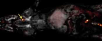
Image: The whole body of a rat can be imaged for blood clots with one PET scan, overlaid here on an MRI image, using the FBP8 probe. The arrow points to a blood clot (Photo courtesy of the American Chemical Society).
The new technique was tested in rats using a probe created by attaching a radionuclide to a peptide. The peptide in the probe binds to the protein fibrin found in blood clots. The radionuclide in the probe can be detected anywhere in a patient’s body using a single Positron Emission Tomography (PET) scan. Researchers experimented with various radionuclides and peptides, to find the brightest PET signal when scanning for blood clots. The best-performing probe that the research team found was Fibrin Binding Probe #8 (FBP8), combined with a copper-64 radionuclide.
The research team from the Athinoula A. Martinos Center for Biomedical Imaging in the Massachusetts General Hospital (MGH; Charlestown, MA, USA) is planning to begin human trials of the probe in fall 2015, but expect that it could take up to five more years of research before the new technique is ready for routine clinical screening.
Peter Caravan, PhD, said, “Patients could end up being scanned multiple times by multiple techniques in order to locate a clot. We sought a method that could detect blood clots anywhere in the body with a single whole-body scan. The probes all had a similar affinity to fibrin in vitro, but in rats, their performances were quite different. The best probe was the one that was the most stable. Of course, the big question is, ‘How well will these perform in patients?’”
Related Links:
Massachusetts General Hospital
American Chemical Society


 Guided Devices.jpg)


