Novel Imaging Technique Could Predict Brain Bleeding Following a Stroke
By MedImaging International staff writers
Posted on 28 Jun 2016
Scientists in the US have used MRI brain scans of stroke patients to confirm the relationship between the extent that the Blood-Brain Barrier (BBB) has been disrupted, and the severity of bleeding, after invasive stroke therapy.Posted on 28 Jun 2016
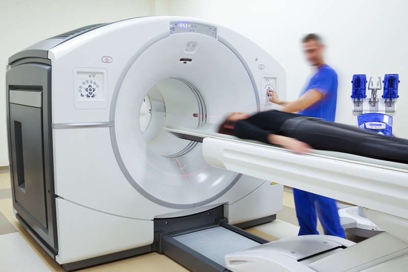
The researchers analyzed Magnetic Resonance Imaging (MRI) scans of more than one hundred stroke patients. The scans were taken before clinicians treated the patients with endovascular therapy, within 12 hours of the onset of the stroke.
.")
Image: The image shows MRI scans of one patient, taken before and after treatment, and reveals the correlation between the area and size of bleeding after treatment, and the disruption to the blood-brain barrier before therapy (Photo courtesy of Dr. Leigh, NINDS).
The study was carried out by scientists at the US National Institutes of Health (NIH) National Institute of Neurological Disorders and Stroke (NINDS) and published online before print on June 17, 2016, in the journal Neurology.
The study authors concluded that the extent of disruption to the brain BBB, as revealed in MRI image scans, could help clinicians determine which patients would be unlikely to benefit from endovascular therapy.
Richard Leigh, M.D., scientist at NINDS, and one of the study authors, said, "The biggest impact of this research is that information from MRI scans routinely collected at a number of research hospitals and stroke centers can inform treating physicians on the risk of bleeding. It is too early to say how these images will be able to help guide clinical decisions, but they can expand how we think about stroke, especially as we try to broaden treatment options for this disease that can have devastating consequences."
Related Links:
National Institute of Neurological Disorders and Stroke

Gold Member
Solid State Kv/Dose Multi-Sensor
AGMS-DM+

New
Ultrasound Table
Ergonomic Advantage (EA) Line

Thyroid Shield
Standard Thyroid Shield

New
Mobile Digital C-arm X-Ray System
HHMC-200D
.jpg)