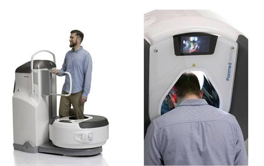
Novel CBCT Offers Orthopedic, Head and Neck Imaging
By MedImaging International staff writers
Posted on 03 Mar 2018
An improved cone beam computerized tomography (CBCT) scanner now offers a three-dimensional (3D) imaging solution for head and neck imaging with enhanced image quality.Posted on 03 Mar 2018
The newest iteration of the Planmed (Helsinki, Finland) Verity scanner has been enhanced to meet all imaging needs within the ear, nose, and throat (ENT) domain, as well as meeting basic dental imaging needs. Verity now includes the Planmeca CALM motion artefact correction, an algorithm that eliminates the need for retakes by cancelling the effects of patient movement, a feature that not only saves time but also guards against unnecessary radiation doses.

Image: An upgraded CBCT scanner now offers head and neck scanning options (Photo courtesy of Planmed).
In addition, the innovative Planmeca ultra low dose (ULD) imaging protocol is now available for the Planmed Verity. Planmeca ULD combines clinical image quality with extremely low effective patient doses, enabling 3D imaging with a dose close to that of standard imaging. The protocol allows reducing the already low effective patient doses by 50% without compromising diagnostic image quality. Finally, new volume positioning possibilities enable enhanced accuracy to the imaging area, thus reducing the amount of radiation patients are exposed to.
“This unit is now more versatile than ever before. It is an easy-to-use and cost-efficient solution making it a great tool for any imaging center, clinic, or hospital,” said Jan Moed, managing director of Planmed. “New innovative features such as Planmeca CALM and Planmeca ULD are great examples of the fruits of our deep collaboration with our parent company Planmeca. I’m extremely excited to see the heights our unique Planmed Verity scanner can reach within the world of medical imaging.”
During CBCT, the region of interest is centered in the field of view. A single 200-degree rotation acquires a volumetric data set, which is used to produce a digital volume composed of 3D voxels of anatomical data that can then be manipulated and visualized. CBCT has only recently become practical with the introduction of large-area high-speed digital X-ray imagers, such as hydrogenated amorphous silicon (a-Si:H) based flat-panel detectors (FPDs).


