New 3D-Printable Materials for Reconstructive Surgery Can Be Monitored Using X-Ray or CT
Posted on 23 Feb 2024
Over the past decade, gelatin-based materials have captured significant attention in research due to their simplicity in production, non-toxic nature, affordability, biodegradability, and crucially, their ability to facilitate cell growth. These attributes make them highly suitable for use in plastic and reconstructive surgery. When a surgeon implants such material into a wound, the body progressively decomposes it and substitutes it with its own tissue. This process not only expedites wound healing but also allows for the reshaping of tissues, such as in breast reconstruction post-mastectomy. Additionally, these materials are valuable for 3D printing custom implants for individual patients. Despite these advantages, there has been a significant challenge: tracking the degradation of these materials in the body has been problematic with traditional imaging techniques.
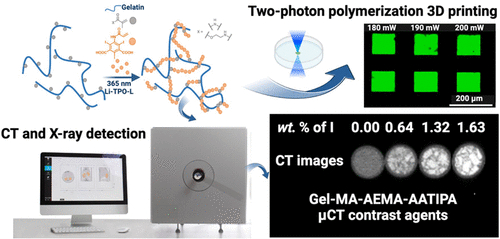
Researchers from IOCB Prague (Prague, Czech Republic) and Ghent University (Ghent, Belgium) have been refining the properties of gelatin-based materials and have introduced 3D-printable materials that are easily trackable using X-ray machines or computed tomography (CT). By incorporating a radiopaque (X-ray-contrast) agent, they have enabled the observation of the rate at which implants shrink and whether they sustain any damage. A whole series of academic papers is being written on this topic. The initial paper introduced a gelatin-based material visible via magnetic resonance imaging (MRI). The most recent publication describes materials that are detectable with X-ray and CT imaging.

This advancement allows for the ongoing monitoring of these implants, observing their biodegradation and identifying potential mechanical failures. Such data are immensely valuable in clinical settings. Leveraging this information, the biodegradation of implants can be customized to align with specific clinical needs. This is vital because tissue growth rates vary across the human body, necessitating the adaptation of implant properties accordingly. The ultimate aim is to align the biodegradation rate of these implants with the growth rate of healthy tissue. In light of these innovations, the two collaborating institutions have filed a joint patent application for the use of these materials in plastic and reconstructive surgery.
Related Links:
IOCB Prague
Ghent University


 Guided Devices.jpg)


