Breakthrough in Nanosized Contrast Agents to Enhance Magnetic Resonance Imaging
Posted on 06 Dec 2023
Magnetic resonance imaging (MRI) plays a vital role in cancer diagnosis by capturing detailed images of soft tissues. For enhanced tumor visibility in MRI scans, doctors often administer contrast agents to patients. These agents impact the response of surrounding hydrogen ions to MRI's radiofrequency pulses. The aim of using contrast agents is to have them specifically accumulate in tumors, thereby heightening their visibility in the scans. However, traditional gadolinium (Gd)-chelate contrast agents have nearly reached their performance peak. One of the main challenges is achieving an optimal dose in the distribution of Gd-chelate in tumors, healthy tissue, and blood without excessive Gd doses.
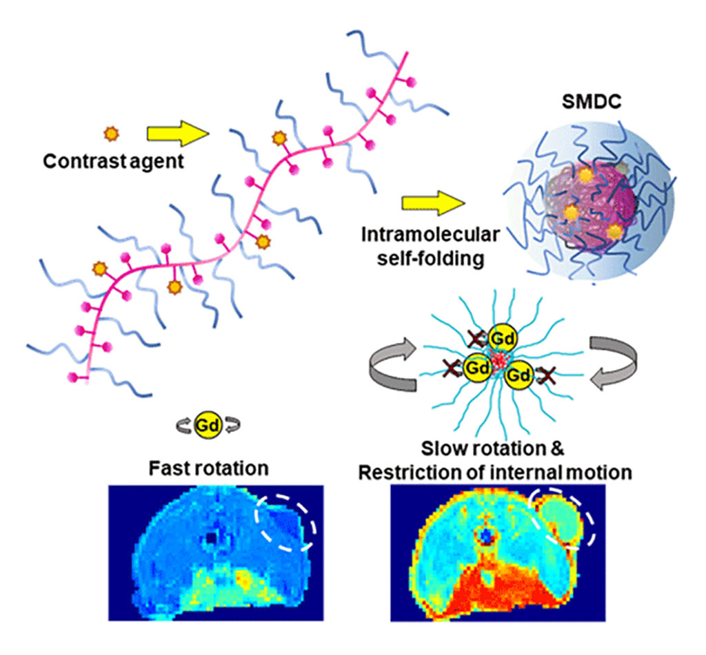
In response, a collaborative study by a research team that included scientists from Tokyo Institute of Technology (Tokyo, Japan) has led to the development of a groundbreaking nano-contrast agent (NCA) using an innovative molecular design. This NCA utilizes Gd as the contrast agent within a "self-folding macromolecular drug carrier (SMDC)." The team integrated clinically approved Gd-containing chelates into a polymer chain comprising poly(ethylene glycol) methyl ether acrylate (PEGA) and benzyl acrylate (BZA). This polymer, featuring both hydrophilic and hydrophobic sections, spontaneously forms a small, capsule-like structure in water, positioning the hydrophobic segments internally and the hydrophilic ones externally.

This methodology allowed for the production of SMDC-Gds molecules under 10 nanometers in size. Experiments on mice with colon cancer demonstrated that these NCAs accumulated more effectively in tumors and were rapidly cleared from the bloodstream, thus enhancing MRI capabilities without toxic side effects. Furthermore, the team uncovered an additional benefit of SMDC-Gds over existing Gd-chelates. The optimal performance of Gd ions is achieved when their movement is restricted, ensuring a consistent and prolonged impact on neighboring hydrogen ions. The SMDC's core/shell structure creates a densely packed molecular environment that limits the rotation and internal motions of Gd ions, thereby resulting in a stronger contrast in MRI images. This will enable the use of alternative elements with safer profiles in patients as well as the environment in the future.
Beyond MRI applications, SMDC-Gds show promise in neutron capture therapy (NCT), an emerging targeted radiotherapy method. In NCT, Gds capture neutrons and emit high-energy radiation, destroying cancer cells in proximity. Experiments showed that NCT with repeated SMDC-Gd injections significantly reduced tumor growth, which can be attributed to the selective accumulation and deep penetration of SMDC-Gds in tumor tissues. The research highlights the potential of SMDCs not just for improved MRI diagnostics, but also as effective cancer treatment modalities and other medical applications.
"This study presents further possibilities for exploiting drug delivery using various therapeutic cargos, and we are currently investigating the development of such SMDC systems," said Associate Professor Yutaka Miura of Tokyo Tech who led the research team.
Related Links:
Tokyo Institute of Technology


