MRI Provides Insights on the Importance of the Placenta
By MedImaging International staff writers
Posted on 10 Jun 2020
A new magnetic resonance imaging (MRI) study reveals differences in blood flow to the placenta in healthy and pre-eclampsia pregnancies.Posted on 10 Jun 2020
Researchers at the University of Nottingham (Nottingham; United Kingdom) conducted an MRI study that scanned 34 women with healthy pregnancies and 13 women diagnosed with preeclampsia in order to explore the function of the human placenta in delivering oxygen to the fetus. The study provided insights on the haemodynamics of the human placenta, and how maternal blood percolates between the villi--which are bathed in a pool of the mother's blood--so that the two different blood supplies are kept separate. They found slow net flow and high net oxygenation were consistent with efficient delivery of oxygen from mother to fetus.

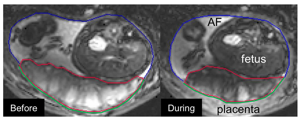
Image: Example of the utero-placental pump; relaxed placenta (L), contracted placenta (R) (Photo courtesy Nelle Dellschaft/ University of Nottingham)
The evidence also substantiated prior hypotheses on the effects of spiral artery remodeling, and also indicated rapid venous drainage from the placenta. The researchers successfully identified Braxton Hicks contractions--which involve the entire uterus--and also a new physiological phenomenon, which they termed the utero-placental pump, in which the placenta and the underlying uterine wall contract independently of the rest of the uterus, expelling maternal blood from the intervillous space. The study was published on May 28, 2020, in PLOS Biology.
“I am part of a team of scientists who have used MRI to look at how blood flows through the placenta to deliver oxygen to the baby. We found that in healthy pregnancies the blood flows very slowly. This seems odd at first, but our other measurements suggest that this is a way in which the placenta can function efficiently,” said lead author Neele Dellschaft, PhD, of the University of Nottingham's School of Physics and Astronomy. “We also found that the normal patterns of flow and oxygenation were much more variable in pre-eclampsia, which can help explain why babies of pre-eclamptic pregnancies tend to be smaller and often have to be delivered before term.”
“At present we have no clinical tools to assess the function of the placenta directly, all we can do is assess the size and growth of the baby and blood flow in the umbilical cord using ultrasound,” said senior author Professor Penny Gowland, PhD, of the Nottingham Biomedical Research Centre. “MRI is hugely effective in providing detailed information of exactly what is happening between the baby and the mother and what is changed in a pre-eclampsia pregnancy. It's also hugely exciting to have discovered a brand new physical phenomenon that takes place during pregnancy.”
Preeclampsia is a complication of pregnancy characterized by hypertension and kidney dysfunction that can cause severe complications for both the mother (including seizures, stroke, renal failure, and liver dysfunction) and the infant (such as low birth weight, preterm delivery, and stillbirth). The condition also increases a woman's risk for cardiovascular disease (CVD) later in life. Currently, there is no cure for preeclampsia, and only childbirth can alleviate the symptoms. An estimated 10 million pregnant women develop it annually, causing approximately 500,000 fetal and neonatal and 76,000 maternal deaths.
Related Links:
University of Nottingham


