MRI Predicts Long-Term Recovery Following Cardiac Arrest
By MedImaging International staff writers
Posted on 30 Oct 2017
Magnetic resonance imaging (MRI) measures of cerebral functional connectivity measured within four weeks of cardiac arrest (CA) are associated with a favorable outcome at one year, according to a new study.Posted on 30 Oct 2017
Researchers at Johns Hopkins University School of Medicine (JHU-SOM; Baltimore, MD, USA), Pitie-Salpetriere Hospital (Paris, France), and other institutions conducted a prospective multicenter study of 46 patients who were comatose after CA in order to assess whether early brain functional connectivity is associated with functional recovery after one year. All participants underwent multiparametric structural and functional MRI about 12 days after CA. Within-network and between-network connectivity was measured in the dorsal attention network (DAN), default-mode network (DMN), salience network (SN), and executive control network (ECN).

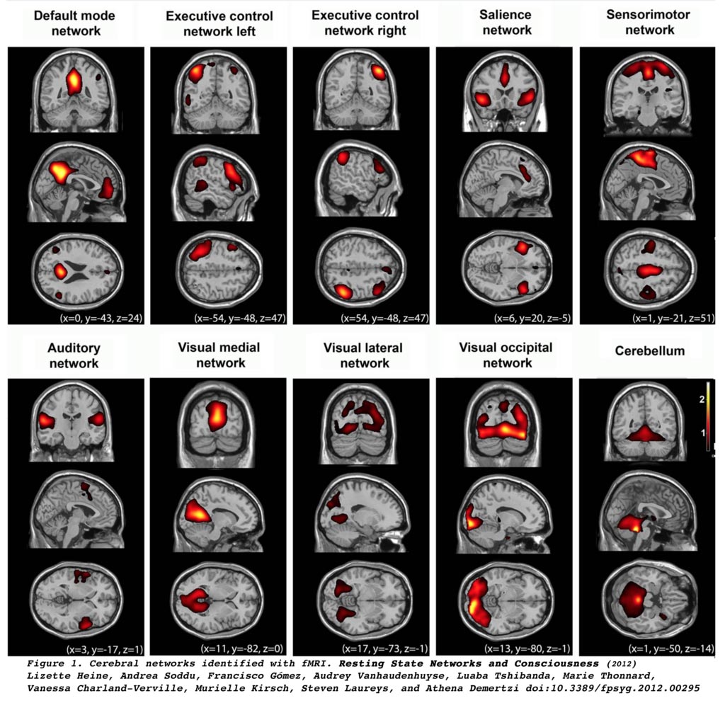
Image: Functional MRI scan shows 10 large-scale brain networks (Photo courtesy of Wikimedia).
The results showed that 11 of the patients had a favorable outcome at one year. Higher within-network DMN connectivity was seen for patients with a favorable outcome, who also had greater anti-correlation between SN and DMN, and between SN and executive control network, when compared with patients with unfavorable outcome; the effect was maintained after adjustment for multiple variables. Compared with fluid-attenuated inversion recovery or diffusion-weighted imaging scores, anti-correlation of SN-DMN predicted outcomes with higher accuracy. The study was published on October 18, 2017, in Radiology.
“Magnetic resonance imaging-based measures of cerebral functional network connectivity obtained in the acute phase of cardiac arrest were independently associated with favorable outcome at one year, warranting validation as early markers of long-term recovery potential in patients with anoxic-ischemic encephalopathy, concluded lead author Haris Sair, MD, and colleagues.
The set of identified brain areas that are linked together in a large-scale network are identified by their function, and provide a coherent framework for understanding cognition. Main networks identified include the DAN, involved in voluntary deployment of attention and reorientation to unexpected events; the DMN, active during introspection; the SN monitors the salience of external inputs and internal brain events; and the ECN is engaged during cognitive tasks that require externally-directed attention, such as working memory, relational integration, response inhibition, and task-set switching.
Related Links:
Johns Hopkins University School of Medicine
Pitie-Salpetriere Hospital


