Technique Boosts the Quality of MRI Scans
By MedImaging International staff writers
Posted on 27 Jun 2017
Researchers have uncovered a new way to boost the quality of Magnetic Resonance Imaging (MRI) brain scans for use in large-scale studies and analyses of stroke outcomes.Posted on 27 Jun 2017
Stroke victims often undergo an MRI brain scan when they reach hospital, but these scans do not have a high enough resolution for research analysis.

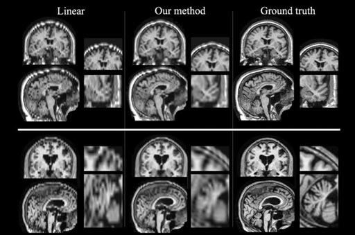
Image: Researchers have devised a new technique to boost the quality of low-resolution MRI scans and make them suitable for use in large-scale studies (Photo courtesy of MIT).
Now a team of researchers at the Massachusetts Institute of Technology (MIT; Cambridge, MA, USA) in collaboration with doctors at the Massachusetts General Hospital (Boston, MA, USA), and many other institutions, has discovered a way to boost the quality of standard MRI scans for future research.
Clinical MRI scans of emergency stroke patients are taken in low-resolution with image slices only every 5-7mm however researchers require higher-resolution images with slices only 1mm apart. To fill in the missing data in the scans from the stroke patients the researchers took information from the entire scan set and used algorithms to recreate the missing anatomical features.
Senior author of the paper, MIT electrical engineering and computer science professor, Polina Golland, said, "These images are quite unique because they are acquired in routine clinical practice when a patient comes in with a stroke. The key idea is to generate an image that is anatomically plausible and to an algorithm looks like one of those research scans, that is completely consistent with clinical images that were acquired. Once you have that, you can apply every state-of-the-art algorithm that was developed for the beautiful research images and run the same analysis, and get the results as if these were the research images."
Related Links:
Massachusetts Institute of Technology
Massachusetts General Hospital


 Guided Devices.jpg)


