First Humans Tested with New MRI Technology
By MedImaging International staff writers
Posted on 19 Jun 2017
A new metamaterial-enhanced MRI technique has been tested for the first time on humans.Posted on 19 Jun 2017
The technique enhances the local sensitivity of Magnetic Resonance Imaging (MRI) scanners and could be used to acquire higher-resolution MRI images, reduce image acquisition time, improve patient comfort, and improve the diagnosis of disease.

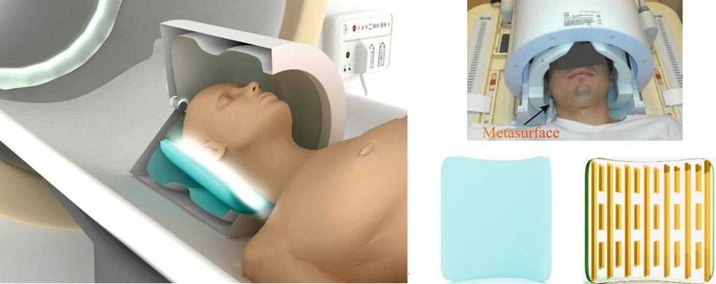
Image: A patient inside an MRI scanner with metasurface-enhanced coil arrays (Photo courtesy of ITMO University).
The new technology was designed and tested by scientists from the Leiden University Medical Center (Leiden, the Netherlands) and ITMO University in (Saint Petersburg, Russia), and was published in the May 2017 issue of the journal Scientific Reports.
Current MRI technology suffers from an intrinsically lower signal-to-noise ratio, and longer acquisition time than Computed Tomography (CT), or ultrasound. However, the new MRI technique enhances local sensitivity and enhances the signal-to-noise ratio. The technique uses a thin metasurface of conducting copper strips attached to a thin flexible substrate, and integrated this into close-fitting receive coil arrays inside an MRI scanner.
Rita Schmidt, researcher at the Department of Radiology, Leiden University Medical Center, and first author of the study, said, "We placed such a metasurface under the patient's head, after that, the local sensitivity increased by 50 percent. This allowed us to obtain higher image and spectroscopic signals from the occipital cortex. Such devices could potentially reduce the duration of MRI studies and improve its comfort for subjects. Our technology can be applied for producing metamaterial-inspired, ultra-thin devices for many different types of MRI scans, but in each case, one should first carry out a series of computer simulations, as we have done in this work. One needs to make sure that the metasurface is appropriately coupled."
Related Links:
Leiden University Medical Center
ITMO University


