Detailed Neural Fingerprint Remains in Human Brain for Decades
By MedImaging International staff writers
Posted on 12 Sep 2016
Researchers have discovered that the primary somatosensory cortex of the human brain keeps a detailed fingerprint of an amputated hand and fingers for decades.Posted on 12 Sep 2016
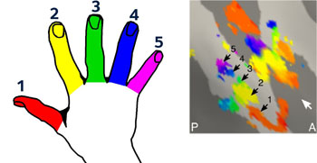
The revelation could change the way the next generation of prosthetic limbs is built. The researchers from universities in the UK and the Netherlands found that the left hand of the amputees showed less brain activity but that the specific patterns of the hand picture fit well to that of a control group that possessed both of their hands.

Image: Research has shown that the human brain keeps a detailed fingerprint of a lost hand and fingers for decades after amputation (Photo courtesy of the University of Oxford).
The study entitled ‘Revealing the neural fingerprints of a missing hand’ was published online, on August 23, 2016, in the open access journal eLIFE. The study was carried out by researchers at the University of Oxford (Oxford, UK) and at other research centers in the UK and the Netherlands.
There were two subjects with amputated hands, and 11 right-handed control subjects. The left hands of amputees had been amputated 25 and 31 years ago respectively, but both subjects still experienced vivid phantom sensations. All subjects underwent a 7-T ultra-high power Magnetic Resonance Imaging (MRI) scan of the primary somatosensory cortex.
Team leader of the study, Dr. Tamar Makin, said: “It has been thought that the hand 'picture' in the brain, located in the primary somatosensory cortex, could only be maintained by regular sensory input from the hand. In fact, textbooks teach that the 'picture' will be 'overwritten' if its primary input stops. If that was the case, people who have undergone hand amputation would show extremely low or no activity related to its original focus in that brain area- in our case, the hand. However, we also know that people experience phantom sensations from amputated body parts, to the extent that someone asked to move a finger can 'feel' that movement. We wanted to look at the information underlying brain activity in phantom movements, to see how it varied from the brain activity of people moving actual hands and fingers.”
Related Links:
University of Oxford


