Restriction Spectrum Imaging with MRI Used to Predict Prostate Cancer Tumor Grade
By MedImaging International staff writers
Posted on 22 Jun 2016
The results of a new study indicate that RSI biomarkers can be used to enhance the ability of MRI to detect prostate cancer tumors, and predict their grade and stage of development, and enable improved treatment options.Posted on 22 Jun 2016
The new imaging tool could help clinicians distinguish between benign or low-grade tumors, and aggressive prostate cancer and guide treatment, and biopsy procedures.

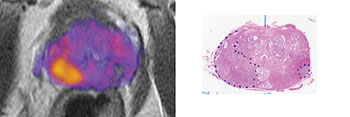
Image: The image on the left is a magnetic resonance image (MRI) of a prostate enhanced with restriction spectrum imaging (RSI). The higher-grade tumor is indicated by orange and yellow. The image on the right is a digitized section of the prostate with the tumors outlined with blue dotted lines (Photo courtesy of UC San Diego Health).
The researchers from the University of California, San Diego School of Medicine (UCSD; San Diego, CA, USA) published the results of the study online in the June 1, 2016, issue of the journal Clinical Cancer Research. According to the researchers Restricted Spectrum Imaging – Magnetic Resonance Imaging (RSI-MRI) can correct for magnetic field distortions.
The researchers enlisted 10 patients for the study and evaluated more than 2,700 discrete data points. The researchers concluded that RSI-MRI could be used as a stand-alone, non-contrast screening tool in the future. An RSI-MRI scan would take only 15 minutes while a standard contrast-enhanced exam today takes between 40 minutes and an hour. Early diagnosis of prostate cancer is crucial to help improve the prognosis of a patient.
Senior author of the study David S. Karow, MD, PhD, assistant professor of radiology at the UCSD, said, “Noninvasive imaging is used to detect disease, but RSI-MRI takes it a step further. We can predict the grade of a tumor sometimes without a biopsy of the prostate tissue. This is taking all that’s good about multi-parametric MRI and making it better. The addition of RSI to a pelvic MRI added between 2.5 to 5 minutes to scanning time making it a fast and highly accurate tool with decreased risk compared to contrast MRI which involves injecting patients with dye.”
Related Links:
University of California, San Diego School of Medicine


