Exclusive Research Partnership to Bring MR Fingerprinting to Clinical Application Stage
By MedImaging International staff writers
Posted on 17 May 2016
A new partnership between a major medical imaging provider and a leading university in the US aims to bring a new Magnetic Resonance (MR) quantitative tissue analysis technique that can identify individual disease tissues, to clinical application.Posted on 17 May 2016
The goal of the researchers is to provide software that can reliably distinguish between healthy and diseased tissue and help identify disease tissues earlier and faster than existing techniques. The MR Fingerprinting (MRF) software package has already been evaluated successfully by several research facilities.

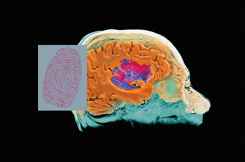
Image: MR Fingerprinting (MRF) can be used to identify individual tissues and diseases quantitatively (Photo courtesy of Siemens Healthcare).
The partnership between Siemens Healthcare (Erlangen, Germany) and Case Western Reserve University (CWRU; Cleveland, OH, USA) was announced in Singapore at the Annual Meeting of the International Society for Magnetic Resonance in Medicine (ISMRM).
MRF provides a non-invasive quantification of tissue properties and can be used to measure multiple parameters simultaneously. The technique provides a unique fingerprint for each type of tissue, disease, or material in the body. MRF can provide a low level of variance across many exam types, different MR scanners, and institutions, and could help clinicians monitor and evaluate patient treatments with greater accuracy.
MRF has previously been used for cardiac examinations and for multiple sclerosis patients. The CWRU research team has successfully used the technique for patients with brain tumors, prostate tumors, and breast cancer patients with liver metastases.
Prof. Siegfried Trattnig from the Medical University of Vienna, who has done initial research with brain tumor and glioma patients, said, "The MR Fingerprint technique lets us see more details than the standard imaging process, and has the potential to redefine MRI. In this way, MRF could help us, as radiologists, to make the paradigm shift from qualitative to quantitative imaging and to incorporate quantitative data into our daily routine."
Related Links:
Siemens Healthcare
Case Western Reserve University


